Deposition Date
2013-11-29
Release Date
2014-04-23
Last Version Date
2024-11-20
Entry Detail
PDB ID:
4NSY
Keywords:
Title:
Wild-type lysobacter enzymogenes lysc endoproteinase covalently inhibited by TLCK
Biological Source:
Source Organism(s):
Lysobacter enzymogenes (Taxon ID: 69)
Expression System(s):
Method Details:
Experimental Method:
Resolution:
1.10 Å
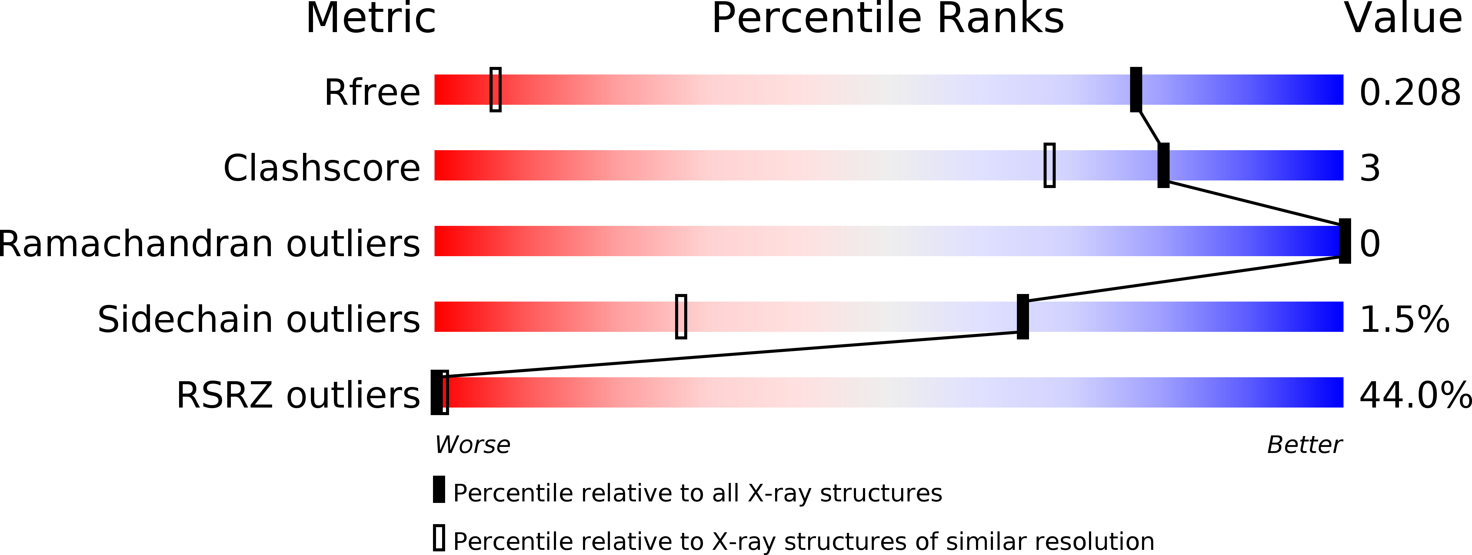
R-Value Free:
0.20
R-Value Work:
0.16
R-Value Observed:
0.16
Space Group:
P 1 21 1