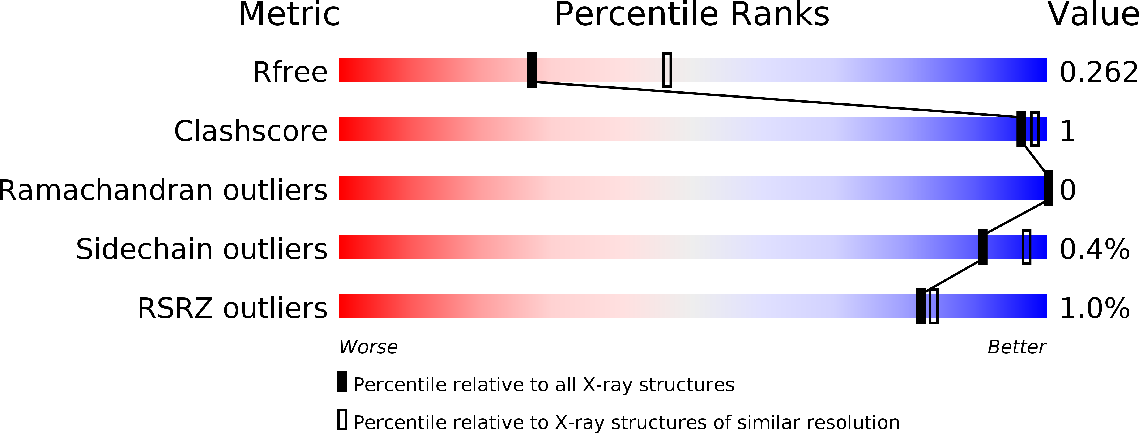
4NON image
Deposition Date 2013-11-20
Release Date 2014-04-02
Last Version Date 2024-02-28
Entry Detail
PDB ID:
4NON
Keywords:
Title:
Crystal structure of GDP-bound A143S mutant of the S. thermophilus FeoB G-domain
Biological Source:
Source Organism:
Host Organism:
Method Details:
Experimental Method:
Resolution:
2.50 Å
R-Value Free:
0.25
R-Value Work:
0.20
R-Value Observed:
0.21
Space Group:
P 1 21 1
Macromolecular Entities
Structures with similar UniProt ID
Protein Blast
Polymer Type:polypeptide(L)
Molecule:Ferrous iron uptake transporter protein B
Gene (Uniprot):feoB
Mutagens:A143S
Chain IDs:A, B
Chain Length:260
Number of Molecules:2
Biological Source:Streptococcus thermophilus
Ligand Molecules
Primary Citation
Structural and functional analysis of a FeoB A143S G5 loop mutant explains the accelerated GDP release rate.
Febs J. 281 2254 2265 (2014)
PMID: 24649829 DOI: 10.1111/febs.12779

Abstact

GTPases (G proteins) hydrolyze the conversion of GTP to GDP and free phosphate, comprising an integral part of prokaryotic and eukaryotic signaling, protein biosynthesis and cell division, as well as membrane transport processes. The G protein cycle is brought to a halt after GTP hydrolysis, and requires the release of GDP before a new cycle can be initiated. For eukaryotic heterotrimeric Gαβγ proteins, the interaction with a membrane-bound G protein-coupled receptor catalyzes the release of GDP from the Gα subunit. Structural and functional studies have implicated one of the nucleotide binding sequence motifs, the G5 motif, as playing an integral part in this release mechanism. Indeed, a Gαs G5 mutant (A366S) was shown to have an accelerated GDP release rate, mimicking a G protein-coupled receptor catalyzed release state. In the present study, we investigate the role of the equivalent residue in the G5 motif (residue A143) in the prokaryotic membrane protein FeoB from Streptococcus thermophilus, which includes an N-terminal soluble G protein domain. The structure of this domain has previously been determined in the apo and GDP-bound states and in the presence of a transition state analogue, revealing conformational changes in the G5 motif. The A143 residue was mutated to a serine and analyzed with respect to changes in GTPase activity, nucleotide release rate, GDP affinity and structural alterations. We conclude that the identity of the residue at this position in the G5 loop plays a key role in the nucleotide release rate by allowing the correct positioning and hydrogen bonding of the nucleotide base.

Legend

Protein

Chemical

Disease

Primary Citation of related structures