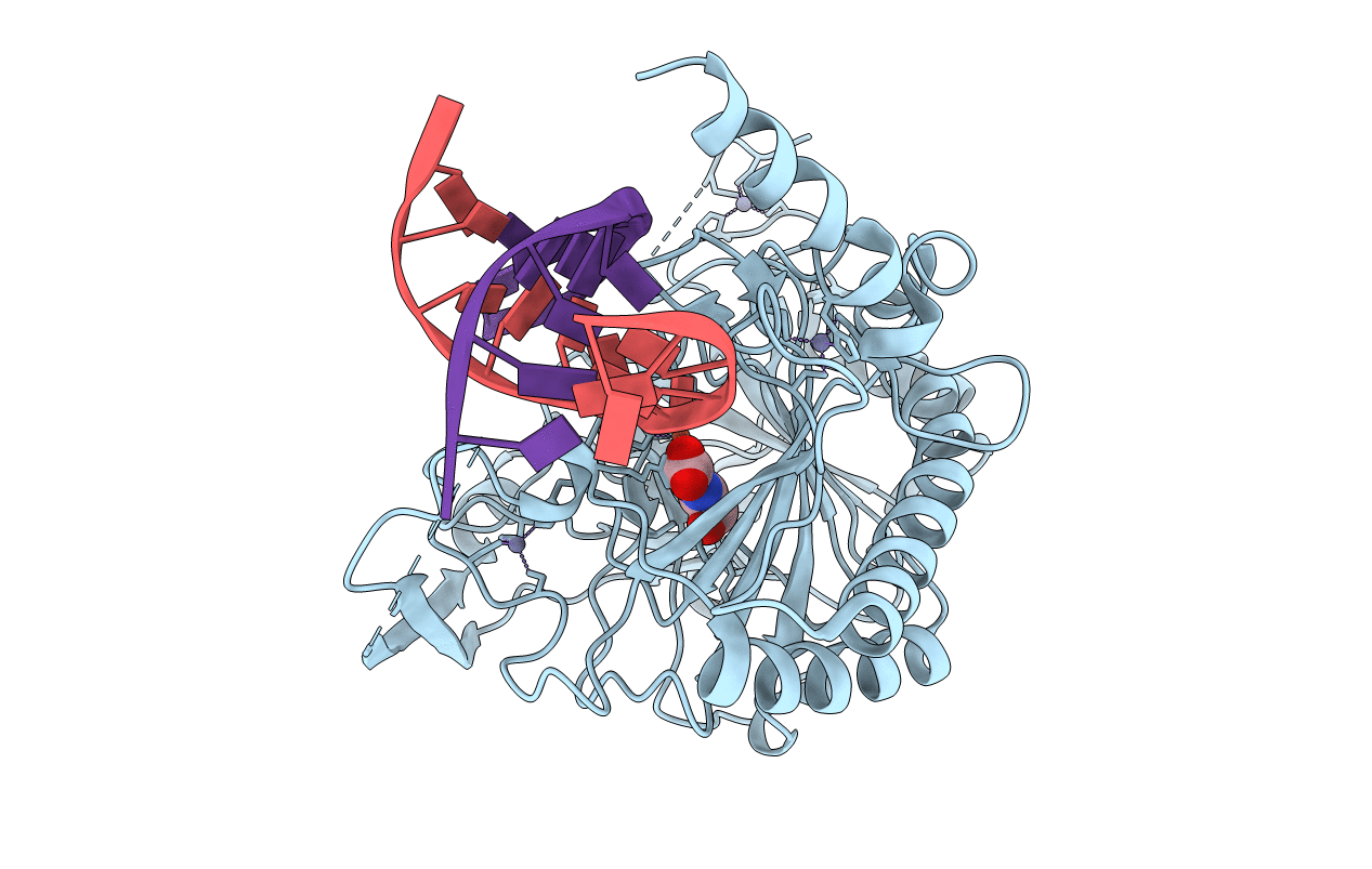
4NM6 image
Entry Detail
PDB ID:
4NM6
Title:
Crystal structure of TET2-DNA complex
Biological Source:
Source Organism:
Host Organism:
PDB Version:
Deposition Date:
2013-11-14
Release Date:
2013-12-18
Method Details:
Experimental Method:
Resolution:
2.03 Å
R-Value Free:
0.24
R-Value Work:
0.20
R-Value Observed:
0.20
Space Group:
C 2 2 21
Macromolecular Entities
Polymer Type:polypeptide(L)
Description:Methylcytosine dioxygenase TET2
Chain IDs:A
Chain Length:463
Number of Molecules:1
Biological Source:Homo sapiens
Modified Residue
Compound ID Chain ID Parent Comp ID Details 2D Image
5CM B DC ?
Primary Citation
Crystal Structure of TET2-DNA Complex: Insight into TET-Mediated 5mC Oxidation.
Cell(Cambridge,Mass.) 155 1545 1555 (2013)
PMID: 24315485 DOI: 10.1016/j.cell.2013.11.020

Abstact

TET proteins oxidize 5-methylcytosine (5mC) on DNA and play important roles in various biological processes. Mutations of TET2 are frequently observed in myeloid malignance. Here, we present the crystal structure of human TET2 bound to methylated DNA at 2.02 Å resolution. The structure shows that two zinc fingers bring the Cys-rich and DSBH domains together to form a compact catalytic domain. The Cys-rich domain stabilizes the DNA above the DSBH core. TET2 specifically recognizes CpG dinucleotide and shows substrate preference for 5mC in a CpG context. 5mC is inserted into the catalytic cavity with the methyl group orientated to catalytic Fe(II) for reaction. The methyl group is not involved in TET2-DNA contacts so that the catalytic cavity allows TET2 to accommodate 5mC derivatives for further oxidation. Mutations of Fe(II)/NOG-chelating, DNA-interacting, and zinc-chelating residues are frequently observed in human cancers. Our studies provide a structural basis for understanding the mechanisms of TET-mediated 5mC oxidation.

Legend

Protein

Chemical

Disease

Primary Citation of related structures