
Deposition Date
2013-11-14
Release Date
2014-03-26
Last Version Date
2023-09-20
Entry Detail
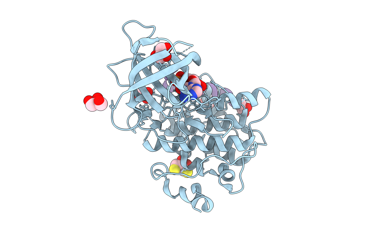
PDB ID:
4NM0
Keywords:
Title:
Crystal structure of peptide inhibitor-free GSK-3/Axin complex
Biological Source:
Source Organism(s):
Homo sapiens (Taxon ID: 9606)
Expression System(s):
Method Details:
Experimental Method:
Resolution:
2.50 Å
R-Value Free:
0.23
R-Value Work:
0.19
R-Value Observed:
0.19
Space Group:
P 61 2 2
