
Deposition Date
2013-11-13
Release Date
2014-06-25
Last Version Date
2024-02-28
Entry Detail
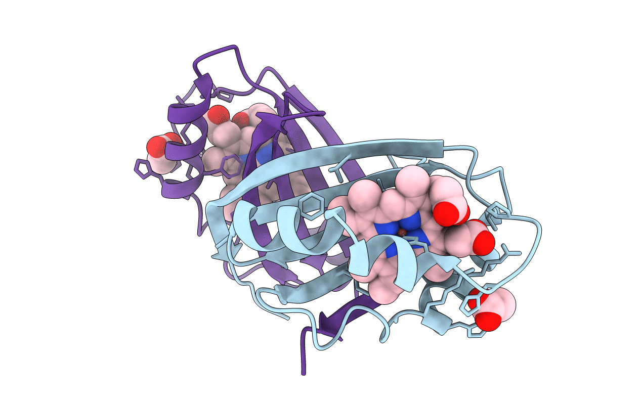
PDB ID:
4NL5
Keywords:
Title:
Mycobacterium tuberculosis heme-degrading protein MhuD in complex with heme and cyanide
Biological Source:
Source Organism(s):
Mycobacterium tuberculosis (Taxon ID: 1773)
Expression System(s):
Method Details:
Experimental Method:
Resolution:
1.90 Å
R-Value Free:
0.22
R-Value Work:
0.17
R-Value Observed:
0.17
Space Group:
P 21 21 21
