Deposition Date
2013-11-10
Release Date
2013-12-25
Last Version Date
2023-09-20
Entry Detail
PDB ID:
4NJK
Keywords:
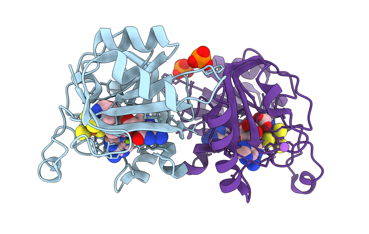
Title:
Crystal Structure of QueE from Burkholderia multivorans in complex with AdoMet, 7-carboxy-7-deazaguanine, and Mg2+
Biological Source:
Source Organism(s):
Burkholderia multivorans (Taxon ID: 395019)
Expression System(s):
Method Details:
Experimental Method:
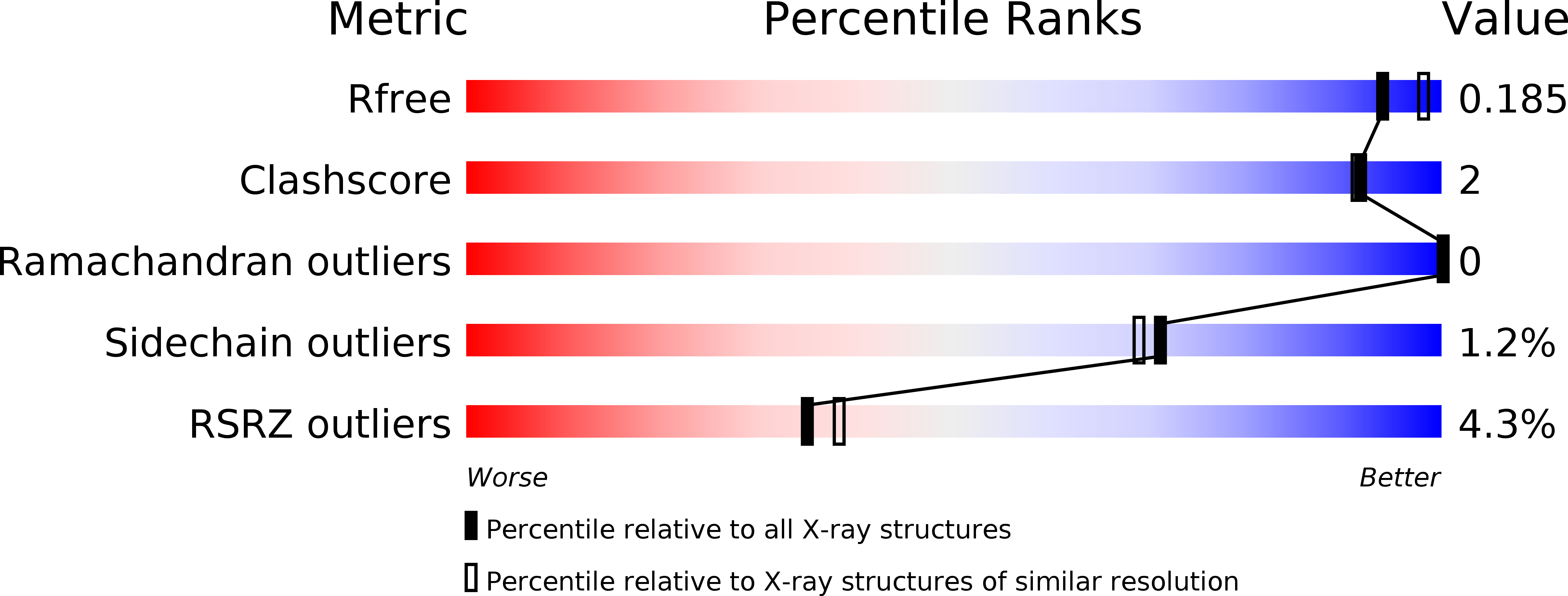
Resolution:
1.91 Å
R-Value Free:
0.18
R-Value Work:
0.15
R-Value Observed:
0.15
Space Group:
P 43 21 2