4NIV image
Deposition Date 2013-11-08
Release Date 2014-02-19
Last Version Date 2024-11-27
Entry Detail
PDB ID:
4NIV
Keywords:
Title:
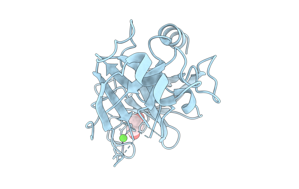
Crystal structure of trypsiligase (K60E/N143H/Y151H/D189K trypsin) trigonal form
Biological Source:
Source Organism:
Bos taurus (Taxon ID: 9913)
Host Organism:
Method Details:
Experimental Method:
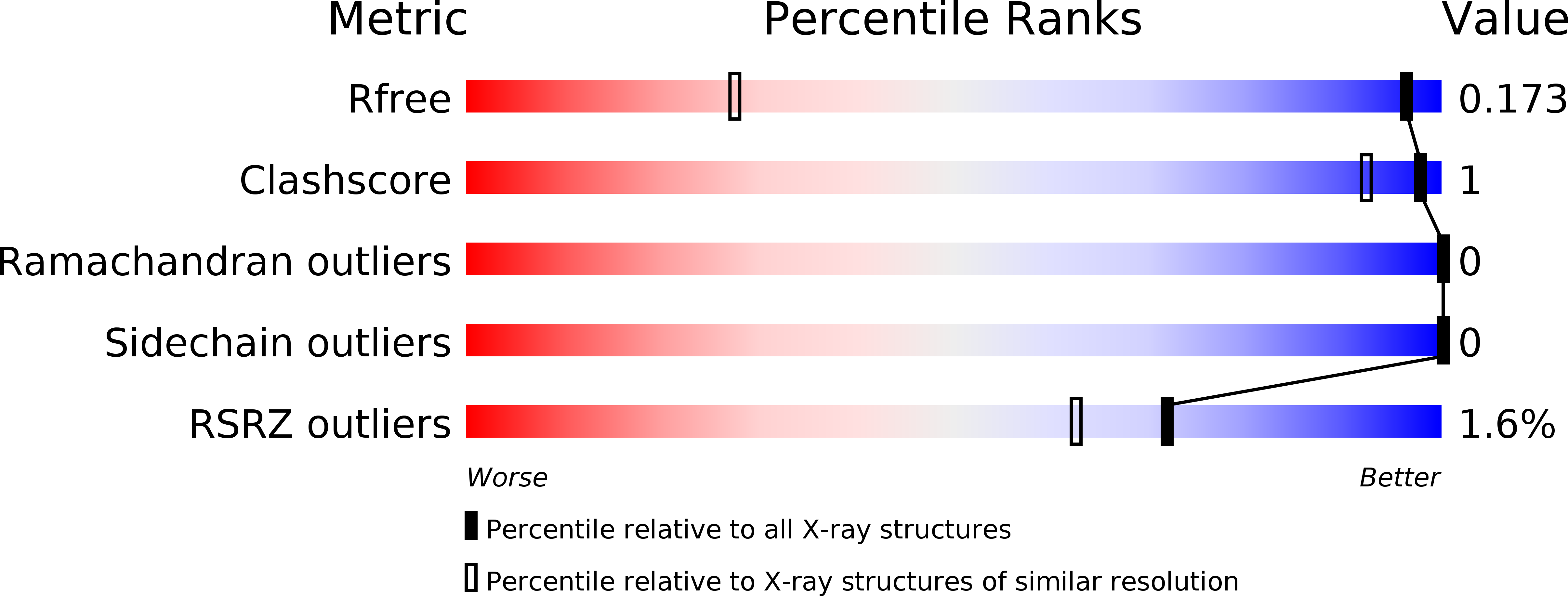
Resolution:
1.00 Å
R-Value Free:
0.17
R-Value Work:
0.15
R-Value Observed:
0.15
Space Group:
P 31 2 1
Macromolecular Entities
Polymer Type:polypeptide(L)
Molecule:Cationic trypsin
Gene (Uniprot):PRSS1
Mutations:K60E, N143H, Y151H, D189K
Chain IDs:A
Chain Length:223
Number of Molecules:1
Biological Source:Bos taurus
Primary Citation
N-terminal protein modification by substrate-activated reverse proteolysis.
Angew.Chem.Int.Ed.Engl. 53 3024 3028 (2014)
PMID: 24520050 DOI: 10.1002/anie.201307736

Abstact

Although site-specific incorporation of artificial functionalities into proteins is an important tool in both basic and applied research, it can be a major challenge to protein chemists. Enzymatic protein modification is an attractive goal due to the inherent regio- and stereoselectivity of enzymes, yet their specificity remains a problem. As a result of the intrinsic reversibility of enzymatic reactions, proteinases can in principle catalyze ligation reactions. While this makes them attractive tools for site-specific protein bioconjugation, competing hydrolysis reactions limits their general use. Here we describe the design and application of a highly specific trypsin variant for the selective modification of N-terminal residues of diverse proteins with various reagents. The modification proceeds quantitatively under native (aqueous) conditions. We show that the variant has a disordered zymogen-like activation domain, effectively suppressing the hydrolysis reaction, which is converted to an active conformation in the presence of appropriate substrates.

Legend

Protein

Chemical

Disease

Primary Citation of related structures