4NEW image
Deposition Date 2013-10-30
Release Date 2014-05-14
Last Version Date 2024-10-16
Entry Detail
PDB ID:
4NEW
Title:
Crystal structure of Trypanothione Reductase from Trypanosoma cruzi in complex with inhibitor EP127 (5-{5-[1-(PYRROLIDIN-1-YL)CYCLOHEXYL]-1,3-THIAZOL-2-YL}-1H-INDOLE)
Biological Source:
Source Organism:
Host Organism:
Method Details:
Experimental Method:
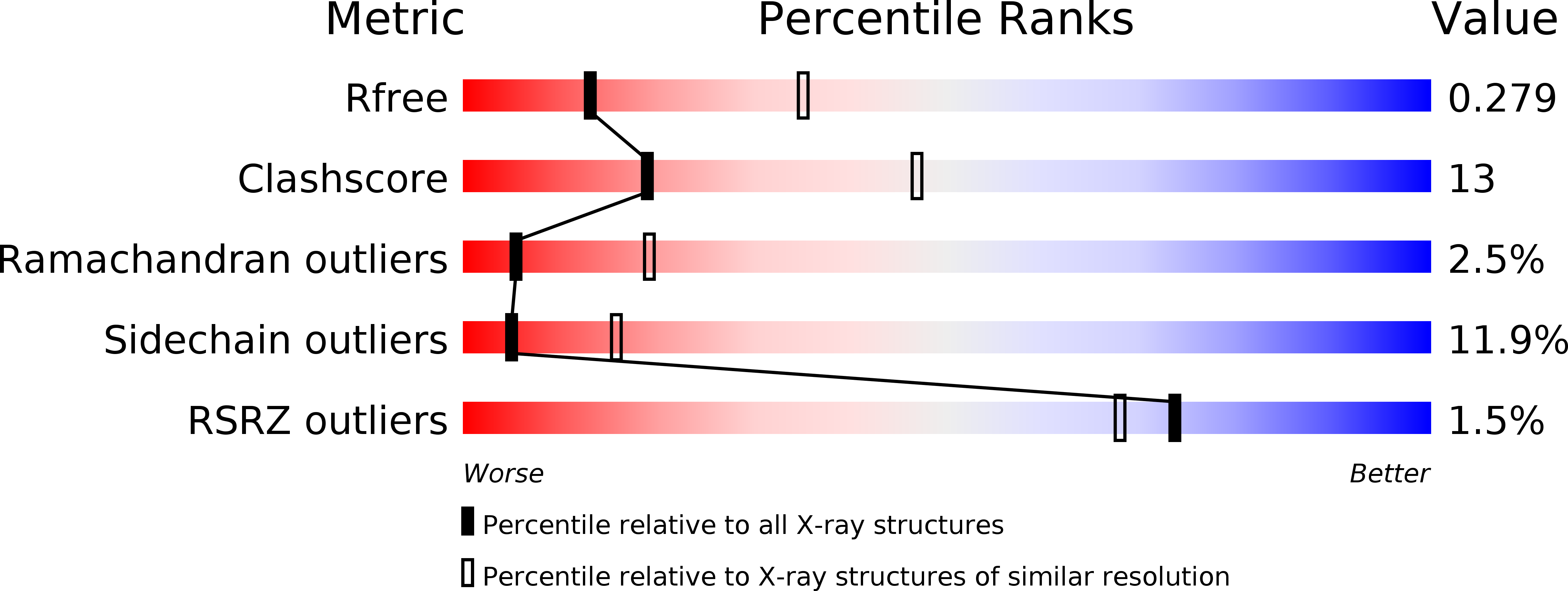
Resolution:
2.80 Å
R-Value Free:
0.27
R-Value Work:
0.23
R-Value Observed:
0.23
Space Group:
P 43 2 2
Macromolecular Entities
Polymer Type:polypeptide(L)
Molecule:Trypanothione reductase, putative
Chain IDs:A
Chain Length:492
Number of Molecules:1
Biological Source:Trypanosoma cruzi
Primary Citation
Binding to large enzyme pockets: small-molecule inhibitors of trypanothione reductase.
Chemmedchem 9 1880 1891 (2014)
PMID: 24788386 DOI: 10.1002/cmdc.201402032

Abstact

The causative agents of the parasitic disease human African trypanosomiasis belong to the family of trypanosomatids. These parasitic protozoa exhibit a unique thiol redox metabolism that is based on the flavoenzyme trypanothione reductase (TR). TR was identified as a potential drug target and features a large active site that allows a multitude of possible ligand orientations, which renders rational structure-based inhibitor design highly challenging. Herein we describe the synthesis, binding properties, and kinetic analysis of a new series of small-molecule inhibitors of TR. The conjunction of biological activities, mutation studies, and virtual ligand docking simulations led to the prediction of a binding mode that was confirmed by crystal structure analysis. The crystal structures revealed that the ligands bind to the hydrophobic wall of the so-called "mepacrine binding site". The binding conformation and potency of the inhibitors varied for TR from Trypanosoma brucei and T. cruzi.

Legend

Protein

Chemical

Disease

Primary Citation of related structures
Feedback Form
Name
Email
Institute
Feedback