4NEQ image
Deposition Date 2013-10-29
Release Date 2014-04-23
Last Version Date 2024-10-30
Entry Detail
PDB ID:
4NEQ
Keywords:
Title:
The structure of UDP-GlcNAc 2-epimerase from Methanocaldococcus jannaschii
Biological Source:
Source Organism:
Host Organism:
Method Details:
Experimental Method:
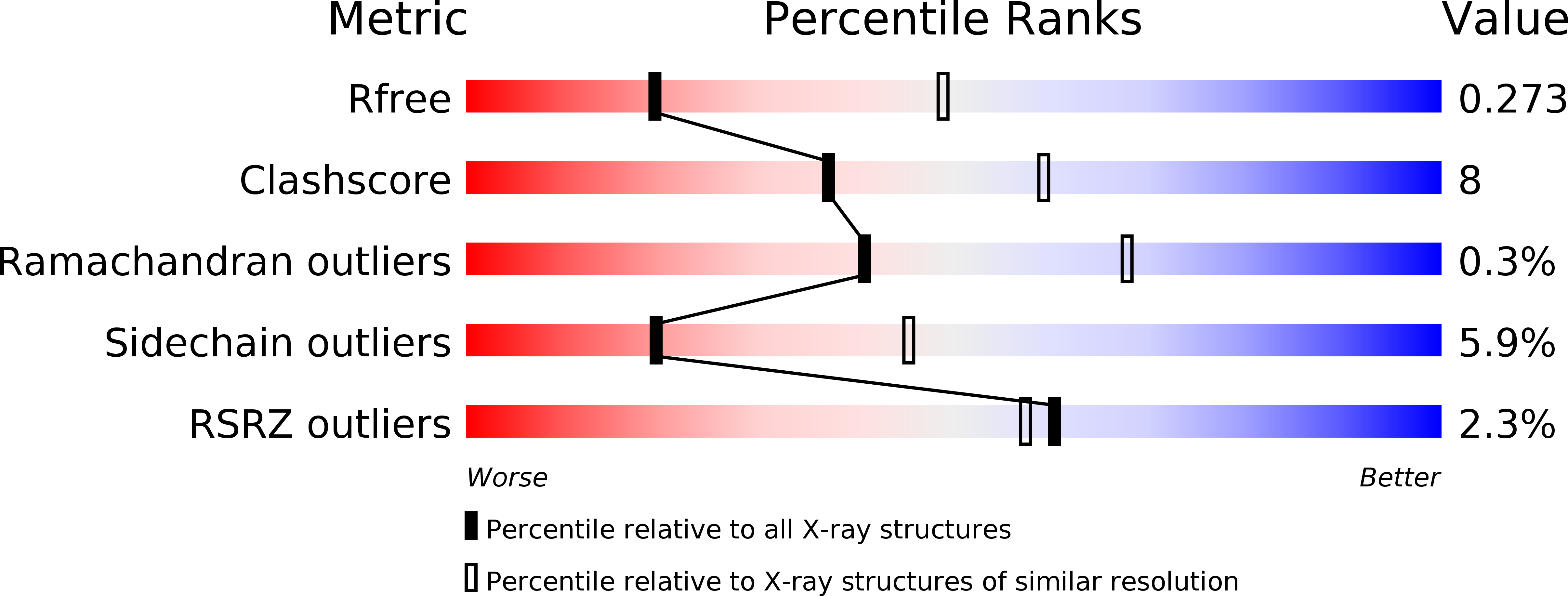
Resolution:
2.85 Å
R-Value Free:
0.27
R-Value Work:
0.19
R-Value Observed:
0.20
Space Group:
P 32 2 1
Macromolecular Entities
Structures with similar UniProt ID
Protein Blast
Polymer Type:polypeptide(L)
Molecule:UDP-N-acetylglucosamine 2-epimerase
Gene (Uniprot):wecB
Chain IDs:A
Chain Length:374
Number of Molecules:1
Biological Source:Methanocaldococcus jannaschii
Modified Residue
Compound ID Chain ID Parent Comp ID Details 2D Image
MSE A MET SELENOMETHIONINE
Ligand Molecules
Primary Citation
Crystal structures of the archaeal UDP-GlcNAc 2-epimerase from Methanocaldococcus jannaschii reveal a conformational change induced by UDP-GlcNAc.
Proteins 82 1519 1526 (2014)
PMID: 24470206 DOI: 10.1002/prot.24516

Abstact

Uridine diphosphate N-acetylglucosamine (UDP-GlcNAc) 2-epimerase catalyzes the interconversion of UDP-GlcNAc to UDP-N-acetylmannosamine (UDP-ManNAc), which is used in the biosynthesis of cell surface polysaccharides in bacteria. Biochemical experiments have demonstrated that mutation of this enzyme causes changes in cell morphology and the thermoresistance of the cell wall. Here, we present the crystal structures of Methanocaldococcus jannaschii UDP-GlcNAc 2-epimerase in open and closed conformations. A comparison of these crystal structures shows that upon UDP and UDP-GlcNAc binding, the enzyme undergoes conformational changes involving a rigid-body movement of the C-terminal domain. We also present the crystal structure of Bacillus subtilis UDP-GlcNAc 2-epimerase in the closed conformation in the presence of UDP and UDP-GlcNAc. Although a structural overlay of these two closed-form structures reveals that the substrate-binding site is evolutionarily conserved, some areas of the allosteric site are distinct between the archaeal and bacterial UDP-GlcNAc 2-epimerases. This is the first report on the crystal structure of archaeal UDP-GlcNAc 2-epimerase, and our results clearly demonstrate the changes between the open and closed conformations of this enzyme.

Legend

Protein

Chemical

Disease

Primary Citation of related structures
Feedback Form
Name
Email
Institute
Feedback