
Deposition Date
2013-10-22
Release Date
2013-12-18
Last Version Date
2024-11-20
Entry Detail
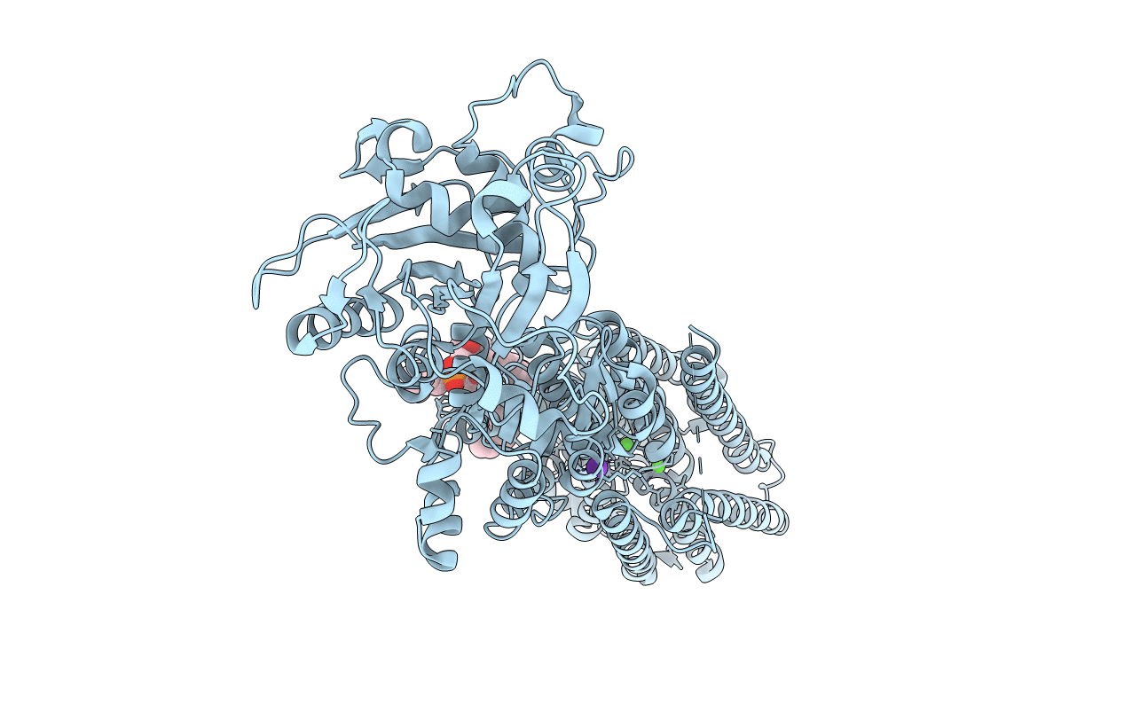
PDB ID:
4NAB
Keywords:
Title:
Structure of the (SR)Ca2+-ATPase mutant E309Q in the Ca2-E1-MgAMPPCP form
Biological Source:
Source Organism(s):
Oryctolagus cuniculus (Taxon ID: 9986)
Expression System(s):
Method Details:
Experimental Method:
Resolution:
3.50 Å
R-Value Free:
0.26
R-Value Work:
0.21
R-Value Observed:
0.21
Space Group:
C 1 2 1
