4NA6 image
Deposition Date 2013-10-21
Release Date 2014-01-29
Last Version Date 2023-09-20
Entry Detail
PDB ID:
4NA6
Keywords:
Title:
Crystal structure of mouse poly(ADP-ribose) glycohydrolase (PARG) catalytic domain mutant E749N
Biological Source:
Source Organism:
Mus musculus (Taxon ID: 10090)
Host Organism:
Method Details:
Experimental Method:
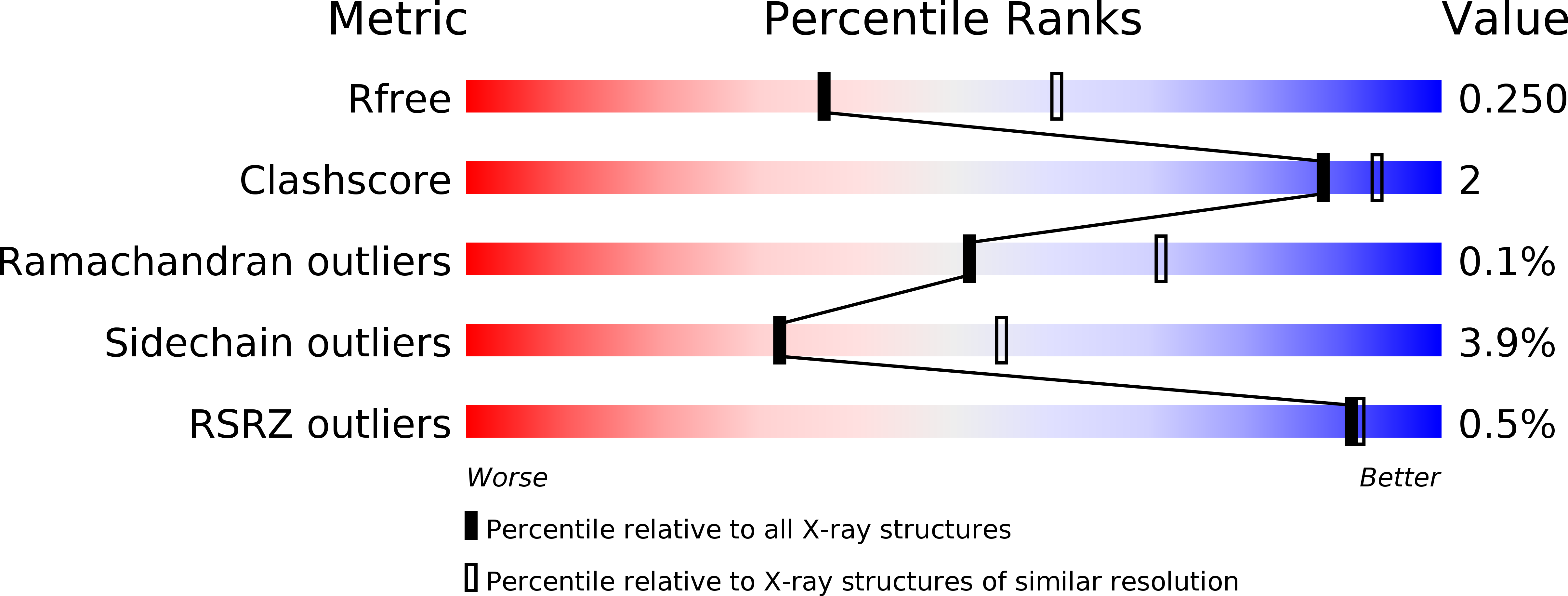
Resolution:
2.48 Å
R-Value Free:
0.25
R-Value Work:
0.21
R-Value Observed:
0.21
Space Group:
P 1 21 1
Macromolecular Entities
Structures with similar UniProt ID
Protein Blast
Polymer Type:polypeptide(L)
Molecule:Poly(ADP-ribose) glycohydrolase
Gene (Uniprot):Parg
Mutagens:E749N
Chain IDs:A, B
Chain Length:522
Number of Molecules:2
Biological Source:Mus musculus
Ligand Molecules
Primary Citation
Crystallographic and biochemical analysis of the mouse poly(ADP-ribose) glycohydrolase.
Plos One 9 e86010 e86010 (2014)
PMID: 24465839 DOI: 10.1371/journal.pone.0086010

Abstact

Protein poly(ADP-ribosyl)ation (PARylation) regulates a number of important cellular processes. Poly(ADP-ribose) glycohydrolase (PARG) is the primary enzyme responsible for hydrolyzing the poly(ADP-ribose) (PAR) polymer in vivo. Here we report crystal structures of the mouse PARG (mPARG) catalytic domain, its complexes with ADP-ribose (ADPr) and a PARG inhibitor ADP-HPD, as well as four PARG catalytic residues mutants. With these structures and biochemical analysis of 20 mPARG mutants, we provide a structural basis for understanding how the PAR polymer is recognized and hydrolyzed by mPARG. The structures and activity complementation experiment also suggest how the N-terminal flexible peptide preceding the PARG catalytic domain may regulate the enzymatic activity of PARG. This study contributes to our understanding of PARG catalytic and regulatory mechanisms as well as the rational design of PARG inhibitors.

Legend

Protein

Chemical

Disease

Primary Citation of related structures