4N8V image
Deposition Date 2013-10-18
Release Date 2014-02-05
Last Version Date 2024-11-06
Entry Detail
PDB ID:
4N8V
Keywords:
Title:
Crystal structure of killer cell immunoglobulin-like receptor KIR2DS2 in complex with HLA-A
Biological Source:
Source Organism:
Homo sapiens (Taxon ID: 9606)
Vaccinia virus WR (Taxon ID: 10254)
Host Organism:
Method Details:
Experimental Method:
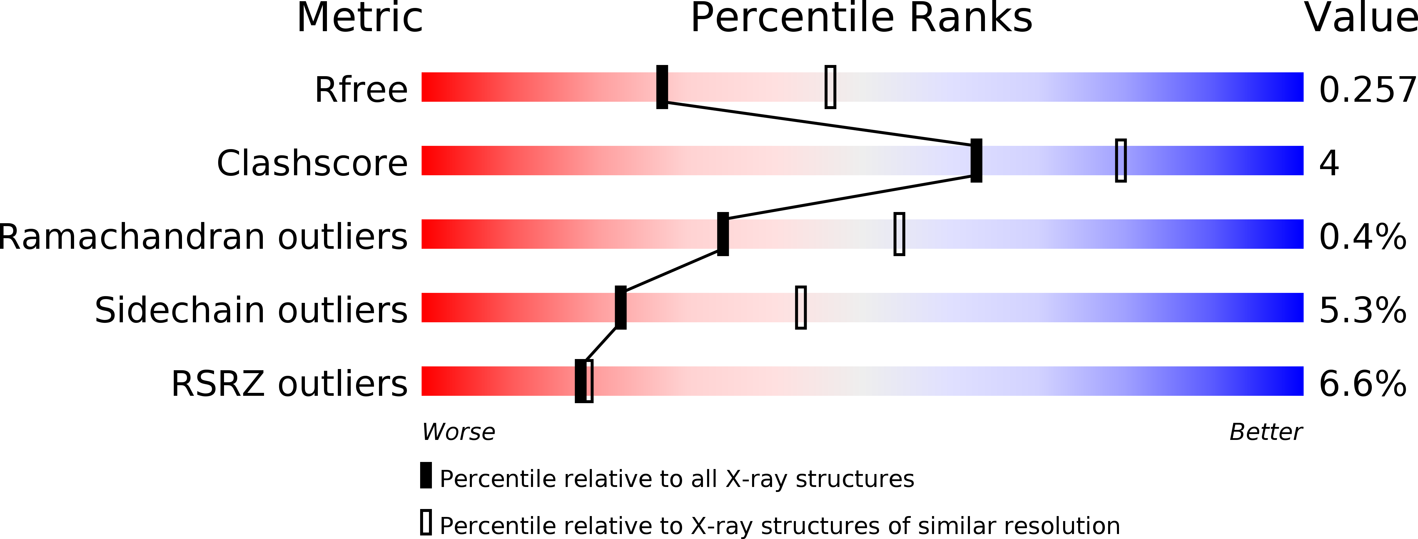
Resolution:
2.50 Å
R-Value Free:
0.25
R-Value Work:
0.22
R-Value Observed:
0.22
Space Group:
P 21 21 21
Macromolecular Entities
Structures with similar UniProt ID
Protein Blast
Polymer Type:polypeptide(L)
Molecule:HLA class I histocompatibility antigen, A-11 alpha chain
Chain IDs:C (auth: A), F (auth: D)
Chain Length:274
Number of Molecules:2
Biological Source:Homo sapiens
Structures with similar UniProt ID
Protein Blast
Polymer Type:polypeptide(L)
Molecule:Beta-2-microglobulin
Gene (Uniprot):B2M
Chain IDs:D (auth: B), G (auth: E)
Chain Length:100
Number of Molecules:2
Biological Source:Homo sapiens
Structures with similar UniProt ID
Protein Blast
Polymer Type:polypeptide(L)
Molecule:peptide from Virion membrane protein A14
Gene (Uniprot):OPG140
Chain IDs:E (auth: C), H (auth: F)
Chain Length:9
Number of Molecules:2
Biological Source:Vaccinia virus WR
Structures with similar UniProt ID
Protein Blast
Polymer Type:polypeptide(L)
Molecule:Killer cell immunoglobulin-like receptor 2DS2
Gene (Uniprot):KIR2DS2
Chain IDs:A (auth: G), B (auth: I)
Chain Length:201
Number of Molecules:2
Biological Source:Homo sapiens
Primary Citation
Activating killer cell immunoglobulin-like receptor 2DS2 binds to HLA-A*11
Proc.Natl.Acad.Sci.USA 111 2662 2667 (2014)
PMID: 24550293 DOI: 10.1073/pnas.1322052111

Abstact

Inhibitory killer cell Ig-like receptors (KIRs) are known to recognize HLA ligands mainly of the HLA-C and Bw4 groups, but the ligands for KIRs are poorly understood. We report here the identification of the cognate ligand for the activating KIR 2DS2 as HLA-A*11:01. The crystal structure of the KIR2DS2-HLA-A*11:01 complex was solved at 2.5-Å resolution and revealed residue-binding characteristics distinct from those of inhibitory KIRs with HLA-C and the critical role of residues Tyr45 and Asp72 in shaping binding specificity to HLA-A*11:01. Using KIR2DS2 tetramers, binding to surface HLA-A*11:01 on live cells was demonstrated and, furthermore, that binding can be altered by residue changes at p8 of the peptide, indicating the influence of peptide sequence on KIR-HLA association. In addition, heteronuclear single quantum coherence NMR was used to map the involvement of critical residues in HLA binding at the interface of KIR and HLA, and validates the data observed in the crystal structure. Our data provide structural evidence of the recognition of A*11:01 by the activating KIR2DS2 and extend our understanding of the KIR-HLA binding spectrum.

Legend

Protein

Chemical

Disease

Primary Citation of related structures
Feedback Form
Name
Email
Institute
Feedback