Deposition Date
2013-10-17
Release Date
2014-05-28
Last Version Date
2024-11-06
Entry Detail
PDB ID:
4N8N
Keywords:
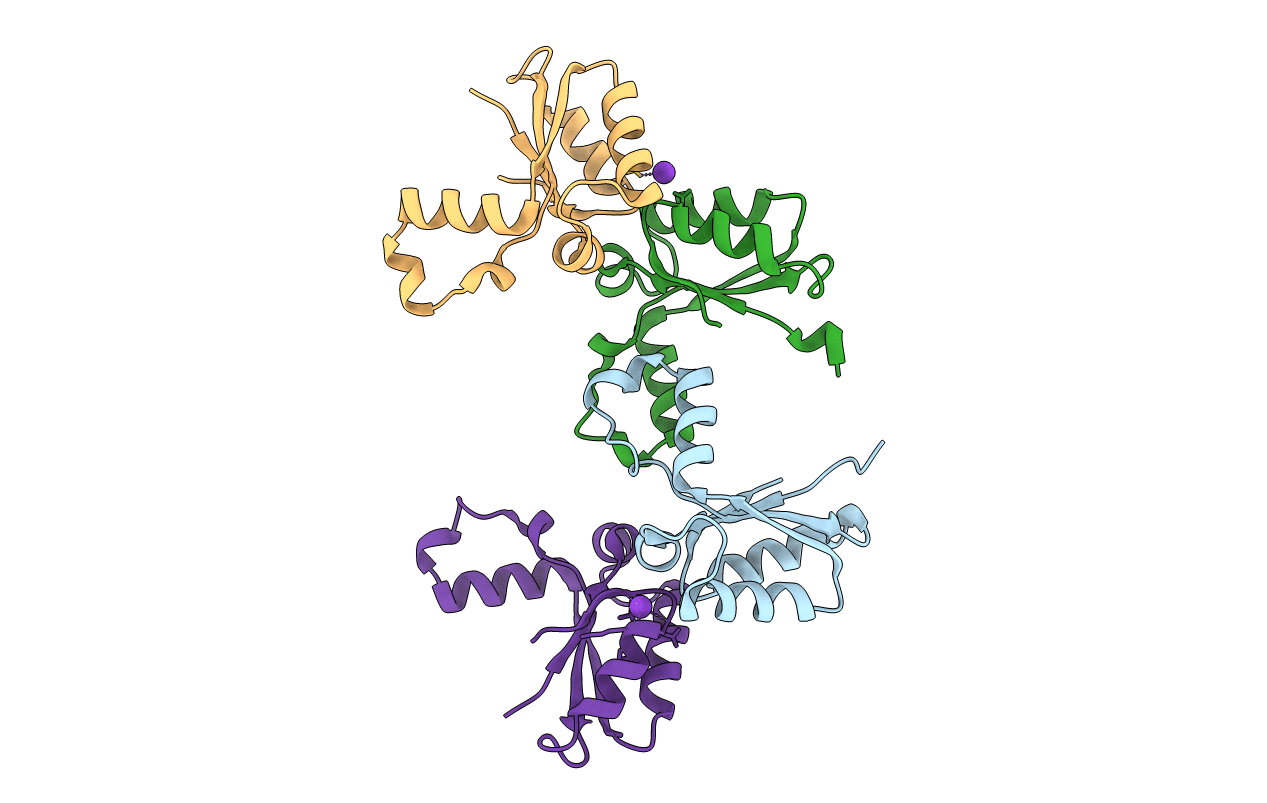
Title:
Crystal structure of Mycobacterial FtsX extracellular domain
Biological Source:
Source Organism(s):
Mycobacterium tuberculosis (Taxon ID: 83332)
Expression System(s):
Method Details:
Experimental Method:
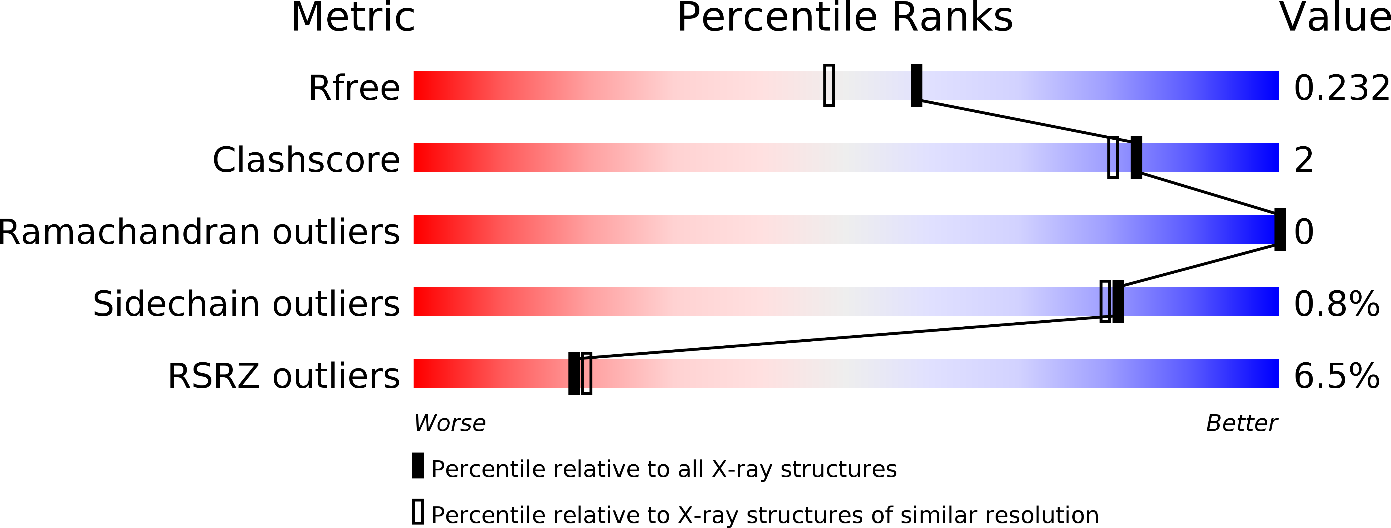
Resolution:
1.87 Å
R-Value Free:
0.23
R-Value Work:
0.18
R-Value Observed:
0.18
Space Group:
P 21 21 2