4N7P image
Entry Detail
PDB ID:
4N7P
Title:
Capturing the haemoglobin allosteric transition in a single crystal form; Crystal structure of half-liganded human haemoglobin without phosphate at 2.8 A resolution.
Biological Source:
Source Organism:
PDB Version:
Deposition Date:
2013-10-16
Release Date:
2014-04-02
Method Details:
Experimental Method:
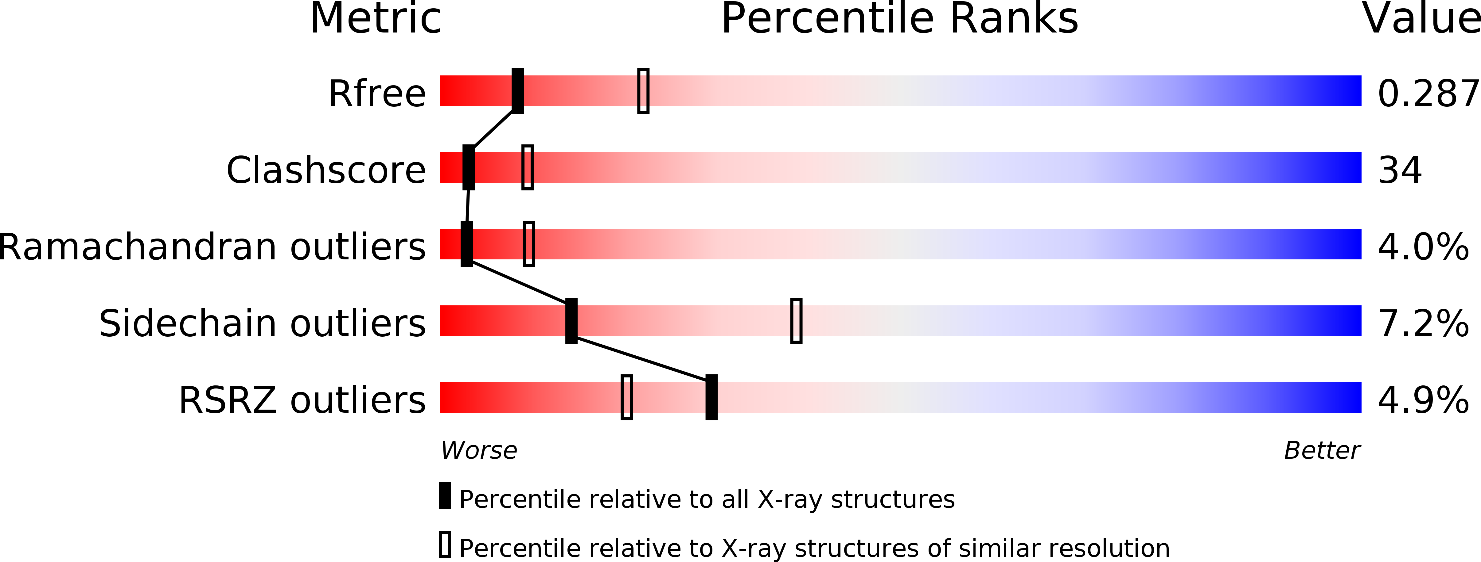
Resolution:
2.81 Å
R-Value Free:
0.28
R-Value Work:
0.25
Space Group:
C 1 2 1
Macromolecular Entities
Polymer Type:polypeptide(L)
Description:Hemoglobin subunit alpha
Chain IDs:A, C, E, G, I, K
Chain Length:141
Number of Molecules:6
Biological Source:Homo sapiens
Polymer Type:polypeptide(L)
Description:Hemoglobin subunit beta
Chain IDs:B, D, F, H, J, L
Chain Length:146
Number of Molecules:6
Biological Source:Homo sapiens
Primary Citation
Capturing the hemoglobin allosteric transition in a single crystal form
J.Am.Chem.Soc. 136 5097 5105 (2014)
PMID: 24635037 DOI: 10.1021/ja500380e

Abstact

Allostery in many oligomeric proteins has been postulated to occur via a ligand-binding-driven conformational transition from the tense (T) to relaxed (R) state, largely on the basis of the knowledge of the structure and function of hemoglobin, the most thoroughly studied of all allosteric proteins. However, a growing body of evidence suggests that hemoglobin possesses a variety of intermediates between the two end states. As such intermediate forms coexist with the end states in dynamic equilibrium and cannot be individually characterized by conventional techniques, very little is known about their properties and functions. Here we present complete structural and functional snapshots of nine equilibrium conformers of human hemoglobin in the half-liganded and fully liganded states by using a novel combination of X-ray diffraction analysis and microspectrophotometric O2 equilibrium measurements on three isomorphous crystals, each capturing three distinct equilibrium conformers. Notably, the conformational set of this crystal form varies according to shifts in the allosteric equilibrium, reflecting the differences in hemoglobin ligation state and crystallization solution conditions. We find that nine snapshot structures cover the complete conformational space of hemoglobin, ranging from T to R2 (the second relaxed quaternary structure) through R, with various relaxed intermediate forms between R and R2. Moreover, we find a previously unidentified intermediate conformer, between T and R, with an intermediate O2 affinity, sought by many research groups over a period of decades. These findings reveal a comprehensive picture of the equilibrium conformers and transition pathway for human hemoglobin.

Legend

Protein

Chemical

Disease

Primary Citation of related structures