4N72 image
Deposition Date 2013-10-14
Release Date 2014-04-23
Last Version Date 2023-09-20
Entry Detail
PDB ID:
4N72
Keywords:
Title:
Catalytic domain from dihydrolipoamide acetyltransferase of pyruvate dehydrogenase from Escherichia coli
Biological Source:
Source Organism:
Host Organism:
Method Details:
Experimental Method:
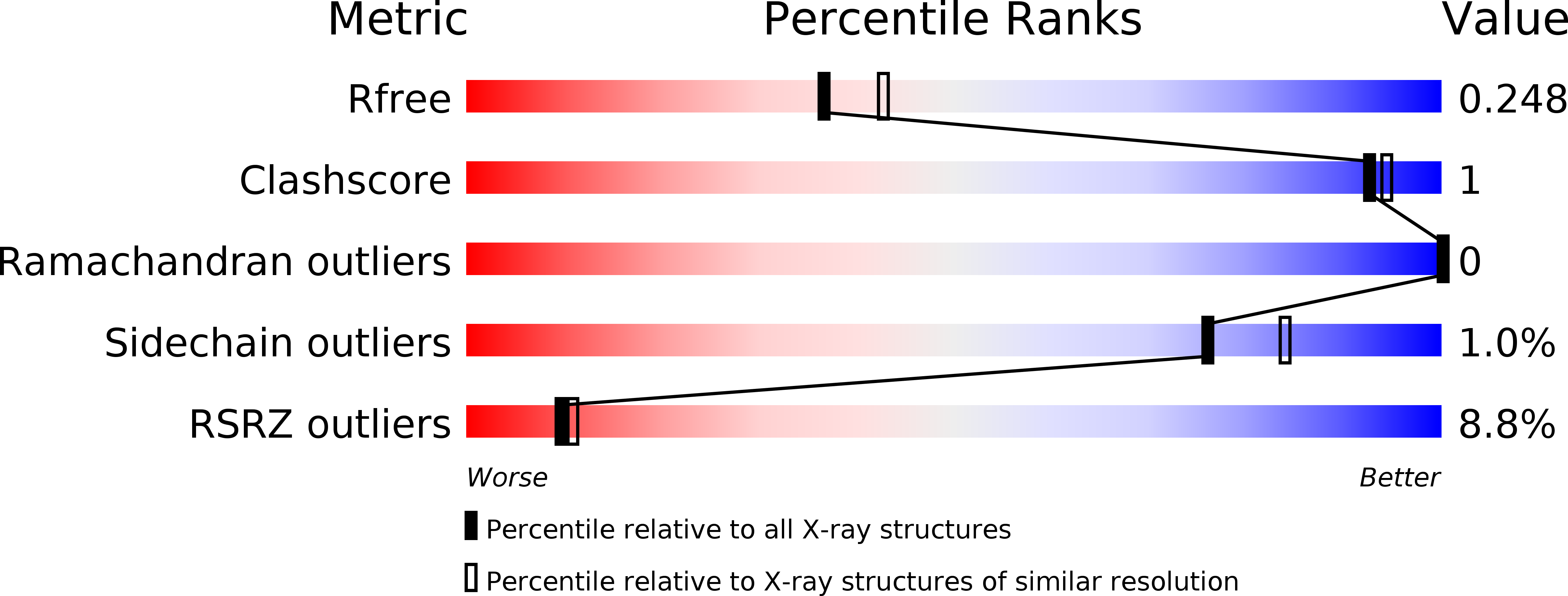
Resolution:
2.25 Å
R-Value Free:
0.24
R-Value Work:
0.20
R-Value Observed:
0.21
Space Group:
P 21 21 21
Macromolecular Entities
Structures with similar UniProt ID
Protein Blast
Polymer Type:polypeptide(L)
Molecule:Pyruvate dehydrogenase (Dihydrolipoyltransacetylase component)
Gene (Uniprot):aceF
Chain IDs:A, B, C
Chain Length:249
Number of Molecules:3
Biological Source:Escherichia coli
Primary Citation
Structure and Function of the Catalytic Domain of the Dihydrolipoyl Acetyltransferase Component in Escherichia coli Pyruvate Dehydrogenase Complex.
J.Biol.Chem. 289 15215 15230 (2014)
PMID: 24742683 DOI: 10.1074/jbc.M113.544080

Abstact

The Escherichia coli pyruvate dehydrogenase complex (PDHc) catalyzing conversion of pyruvate to acetyl-CoA comprises three components: E1p, E2p, and E3. The E2p is the five-domain core component, consisting of three tandem lipoyl domains (LDs), a peripheral subunit binding domain (PSBD), and a catalytic domain (E2pCD). Herein are reported the following. 1) The x-ray structure of E2pCD revealed both intra- and intertrimer interactions, similar to those reported for other E2pCDs. 2) Reconstitution of recombinant LD and E2pCD with E1p and E3p into PDHc could maintain at least 6.4% activity (NADH production), confirming the functional competence of the E2pCD and active center coupling among E1p, LD, E2pCD, and E3 even in the absence of PSBD and of a covalent link between domains within E2p. 3) Direct acetyl transfer between LD and coenzyme A catalyzed by E2pCD was observed with a rate constant of 199 s(-1), comparable with the rate of NADH production in the PDHc reaction. Hence, neither reductive acetylation of E2p nor acetyl transfer within E2p is rate-limiting. 4) An unprecedented finding is that although no interaction could be detected between E1p and E2pCD by itself, a domain-induced interaction was identified on E1p active centers upon assembly with E2p and C-terminally truncated E2p proteins by hydrogen/deuterium exchange mass spectrometry. The inclusion of each additional domain of E2p strengthened the interaction with E1p, and the interaction was strongest with intact E2p. E2p domain-induced changes at the E1p active site were also manifested by the appearance of a circular dichroism band characteristic of the canonical 4'-aminopyrimidine tautomer of bound thiamin diphosphate (AP).

Legend

Protein

Chemical

Disease

Primary Citation of related structures