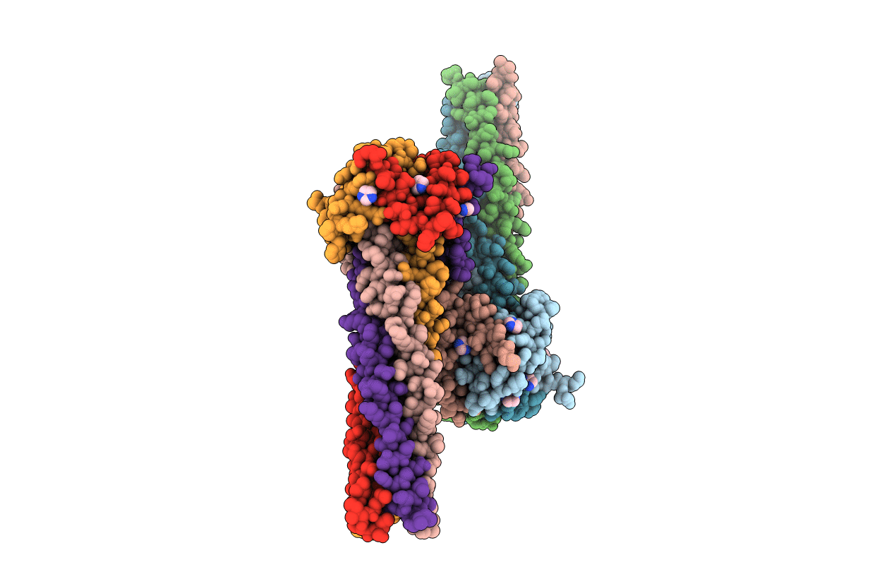
4N5B image
Deposition Date 2013-10-09
Release Date 2013-11-27
Last Version Date 2023-09-20
Entry Detail
PDB ID:
4N5B
Keywords:
Title:
Crystal structure of the Nipah virus phosphoprotein tetramerization domain
Biological Source:
Source Organism:
Nipah virus (Taxon ID: 121791)
Host Organism:
Method Details:
Experimental Method:
Resolution:
2.20 Å
R-Value Free:
0.23
R-Value Work:
0.18
R-Value Observed:
0.18
Space Group:
P 1
Macromolecular Entities
Polymer Type:polypeptide(L)
Molecule:Phosphoprotein
Gene (Uniprot):P/V/W/C
Chain IDs:A, B (auth: E), C (auth: D), D (auth: C), E (auth: H), F, G (auth: B), H (auth: G)
Chain Length:110
Number of Molecules:8
Biological Source:Nipah virus
Primary Citation
Crystal structure of the nipah virus phosphoprotein tetramerization domain.
J.Virol. 88 758 762 (2014)
PMID: 24155387 DOI: 10.1128/JVI.02294-13

Abstact

The Nipah virus phosphoprotein (P) is multimeric and tethers the viral polymerase to the nucleocapsid. We present the crystal structure of the multimerization domain of Nipah virus P: a long, parallel, tetrameric, coiled coil with a small, α-helical cap structure. Across the paramyxoviruses, these domains share little sequence identity yet are similar in length and structural organization, suggesting a common requirement for scaffolding or spatial organization of the functions of P in the virus life cycle.

Legend

Protein

Chemical

Disease

Primary Citation of related structures