Deposition Date
2013-10-02
Release Date
2014-02-05
Last Version Date
2024-10-30
Entry Detail
PDB ID:
4N0U
Keywords:
Title:
Ternary complex between Neonatal Fc receptor, serum albumin and Fc
Biological Source:
Source Organism(s):
Homo sapiens (Taxon ID: 9606)
Expression System(s):
Method Details:
Experimental Method:
Resolution:
3.80 Å
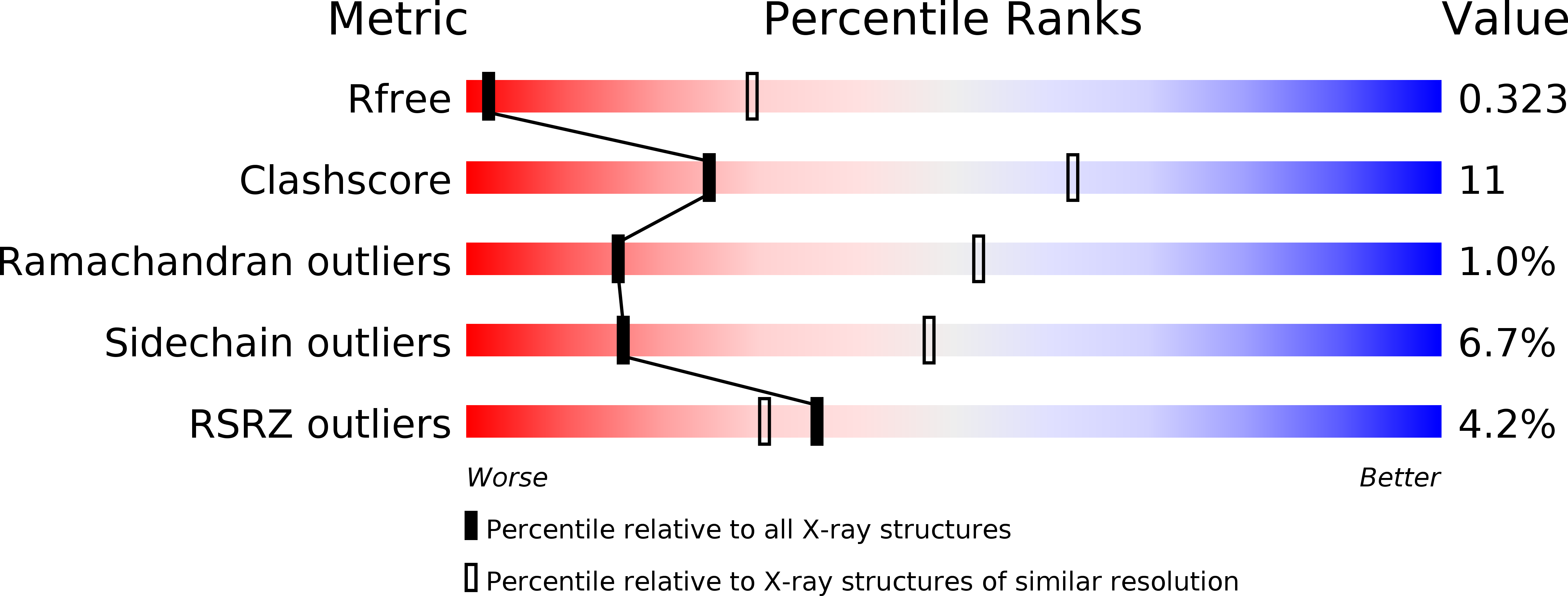
R-Value Free:
0.30
R-Value Work:
0.28
R-Value Observed:
0.28
Space Group:
P 41 21 2