4N08 image
Deposition Date 2013-10-01
Release Date 2014-01-15
Last Version Date 2024-02-28
Entry Detail
PDB ID:
4N08
Keywords:
Title:
Structure of Trypanosoma brucei brucei adenosine kinase (apo)
Biological Source:
Source Organism:
Host Organism:
Method Details:
Experimental Method:
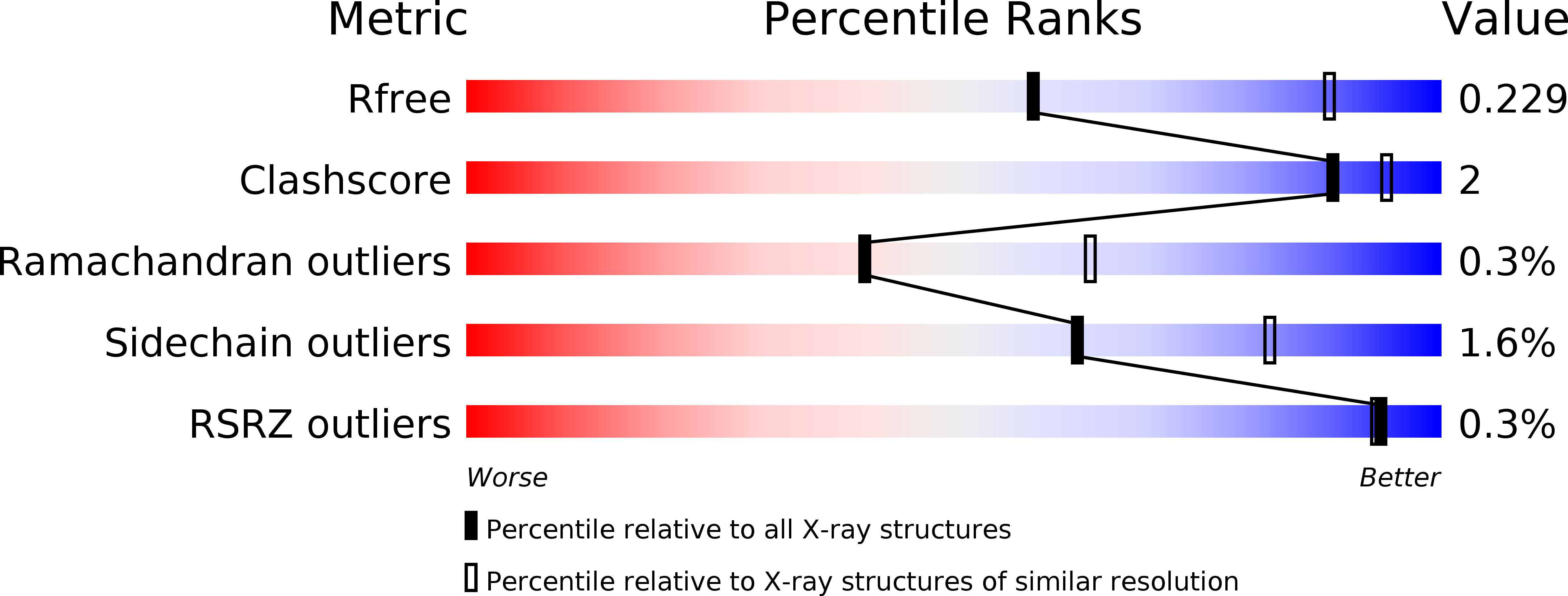
Resolution:
2.60 Å
R-Value Free:
0.23
R-Value Work:
0.16
R-Value Observed:
0.16
Space Group:
P 41 21 2
Macromolecular Entities
Polymer Type:polypeptide(L)
Molecule:Adenosine kinase
Gene (Uniprot):Tb06.4M18.780
Chain IDs:A
Chain Length:348
Number of Molecules:1
Biological Source:Trypanosoma brucei brucei
Primary Citation
Structures of adenosine kinase from Trypanosoma brucei brucei.
Acta Crystallogr F Struct Biol Commun 70 34 39 (2014)
PMID: 24419613 DOI: 10.1107/S2053230X13033621

Abstact

Trypanosoma brucei is a single-cellular parasite of the genus Kinetoplastida and is the causative agent of African sleeping sickness in humans. Adenosine kinase is a key enzyme in the purine-salvage pathway, phosphorylating adenosine to AMP, and also activates cytotoxic analogues such as cordycepin and Ara-A by their phosphorylation. The structures of T. brucei brucei adenosine kinase (TbAK) in its unliganded open conformation and complexed with adenosine and ADP in the closed conformation are both reported to 2.6 Å resolution. The structures give insight into the binding mode of the substrates and the conformational change induced upon substrate binding. This information can be used to guide the improvement of cytotoxic substrate analogues as potential antitrypanosomal drugs.

Legend

Protein

Chemical

Disease

Primary Citation of related structures