4MYC image
Deposition Date 2013-09-27
Release Date 2014-03-26
Last Version Date 2024-02-28
Entry Detail
PDB ID:
4MYC
Title:
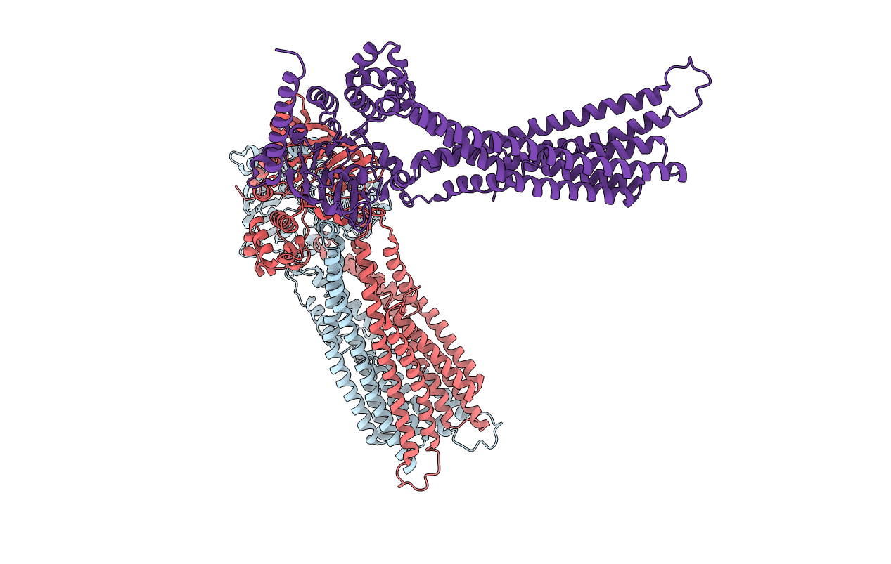
Structure of the mitochondrial ABC transporter, Atm1
Biological Source:
Source Organism(s):
Expression System(s):
Method Details:
Experimental Method:
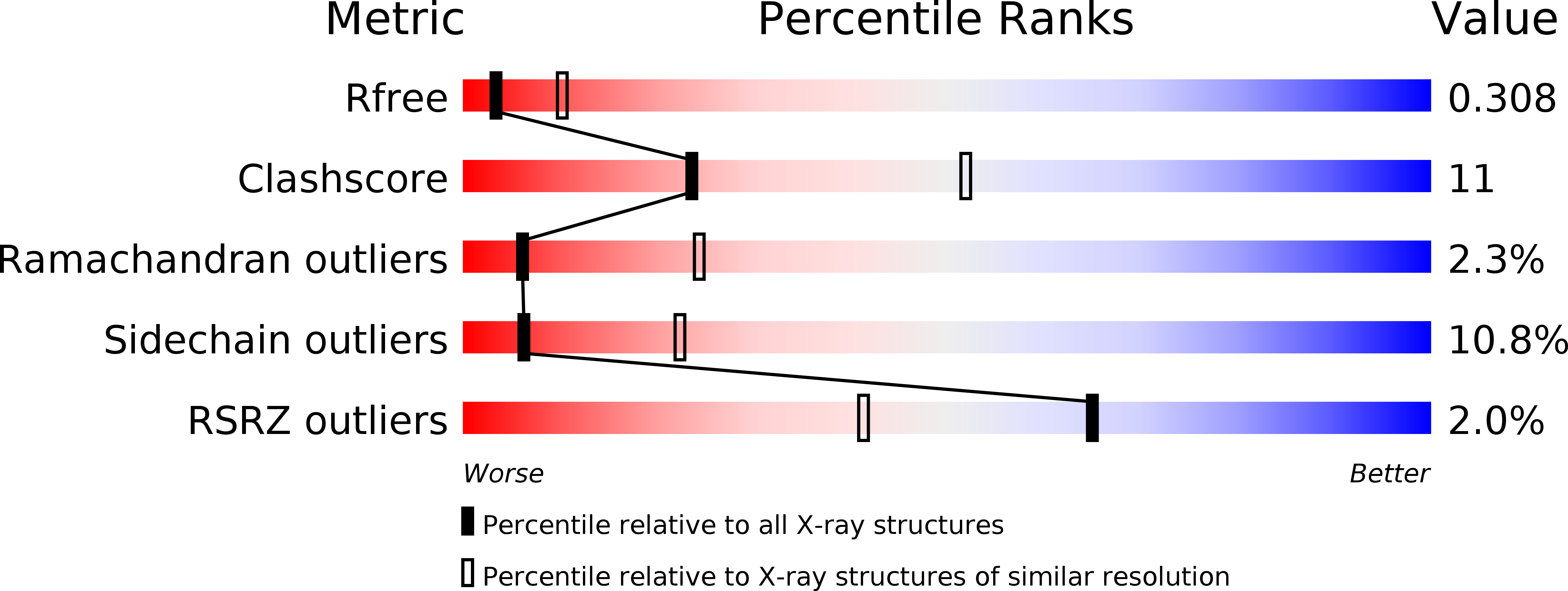
Resolution:
3.06 Å
R-Value Free:
0.29
R-Value Work:
0.25
R-Value Observed:
0.25
Space Group:
P 65 2 2
Macromolecular Entities
Structures with similar UniProt ID
Protein Blast
Polymer Type:polypeptide(L)
Molecule:Iron-sulfur clusters transporter ATM1, mitochondrial
Gene (Uniprot):ATM1
Chain IDs:A, B (auth: C), C (auth: B)
Chain Length:598
Number of Molecules:3
Biological Source:Saccharomyces cerevisiae
Ligand Molecules
Primary Citation
Crystal structures of nucleotide-free and glutathione-bound mitochondrial ABC transporter Atm1.
Science 343 1137 1140 (2014)
PMID: 24604199 DOI: 10.1126/science.1246729

Abstact

The yeast mitochondrial ABC transporter Atm1, in concert with glutathione, functions in the export of a substrate required for cytosolic-nuclear iron-sulfur protein biogenesis and cellular iron regulation. Defects in the human ortholog ABCB7 cause the sideroblastic anemia XLSA/A. Here, we report the crystal structures of free and glutathione-bound Atm1 in inward-facing, open conformations at 3.06- and 3.38-angstrom resolution, respectively. The glutathione binding site includes a residue mutated in XLSA/A and is located close to the inner membrane surface in a large cavity. The two nucleotide-free adenosine 5'-triphosphate binding domains do not interact yet are kept in close vicinity through tight interaction of the two C-terminal α-helices of the Atm1 dimer. The resulting protein stabilization may be a common structural feature of all ABC exporters.

Legend

Protein

Chemical

Disease

Primary Citation of related structures
Feedback Form
Name
Email
Institute
Feedback