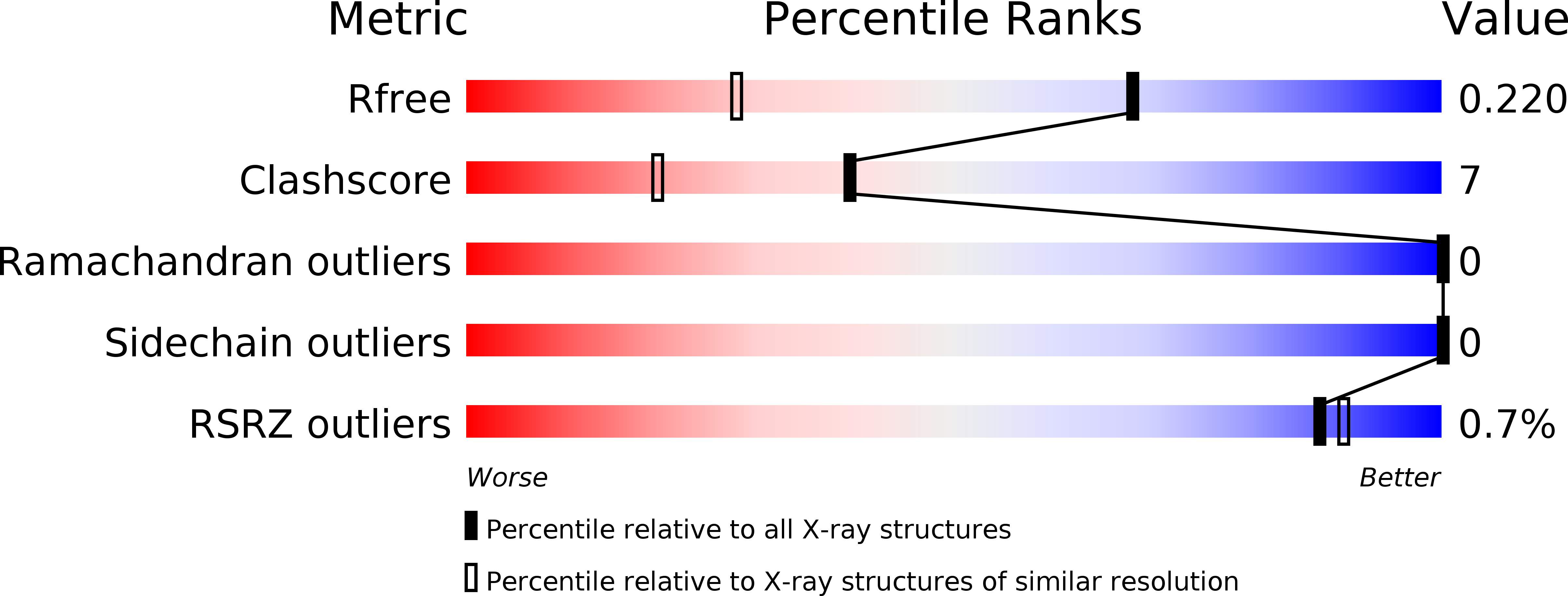
4MXK image
Deposition Date 2013-09-26
Release Date 2014-02-12
Last Version Date 2024-02-28
Entry Detail
PDB ID:
4MXK
Title:
X-ray structure of Fe(II)-ZnPIXFeBMb1
Biological Source:
Source Organism:
Host Organism:
Method Details:
Experimental Method:
Resolution:
1.52 Å
R-Value Free:
0.21
R-Value Work:
0.17
R-Value Observed:
0.18
Space Group:
P 21 21 21
Macromolecular Entities
Structures with similar UniProt ID
Protein Blast
Polymer Type:polypeptide(L)
Molecule:Myoglobin
Gene (Uniprot):MB
Mutagens:L29H, F43H, V68E
Chain IDs:A
Chain Length:153
Number of Molecules:1
Biological Source:Physeter catodon
Primary Citation
Spectroscopic and computational study of a nonheme iron nitrosyl center in a biosynthetic model of nitric oxide reductase.
Angew.Chem.Int.Ed.Engl. 53 2417 2421 (2014)
PMID: 24481708 DOI: 10.1002/anie.201308431

Abstact

A major barrier to understanding the mechanism of nitric oxide reductases (NORs) is the lack of a selective probe of NO binding to the nonheme FeB center. By replacing the heme in a biosynthetic model of NORs, which structurally and functionally mimics NORs, with isostructural ZnPP, the electronic structure and functional properties of the FeB nitrosyl complex was probed. This approach allowed observation of the first S=3/2 nonheme {FeNO}(7) complex in a protein-based model system of NOR. Detailed spectroscopic and computational studies show that the electronic state of the {FeNO}(7) complex is best described as a high spin ferrous iron (S=2) antiferromagnetically coupled to an NO radical (S=1/2) [Fe(2+)-NO(.)]. The radical nature of the FeB -bound NO would facilitate N-N bond formation by radical coupling with the heme-bound NO. This finding, therefore, supports the proposed trans mechanism of NO reduction by NORs.

Legend

Protein

Chemical

Disease

Primary Citation of related structures