4MX3 image
Deposition Date 2013-09-25
Release Date 2014-01-15
Last Version Date 2023-09-20
Entry Detail
PDB ID:
4MX3
Title:
Crystal Structure of PKA RIalpha Homodimer
Biological Source:
Source Organism:
Bos taurus (Taxon ID: 9913)
Host Organism:
Method Details:
Experimental Method:
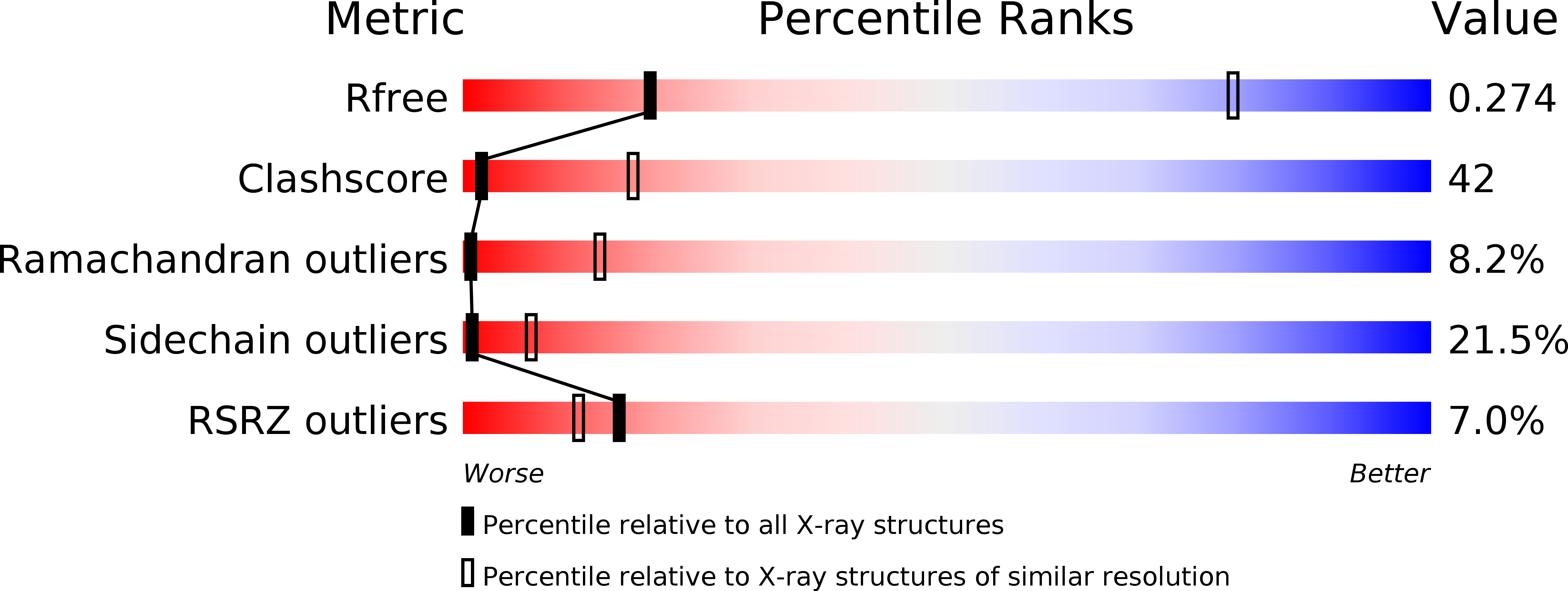
Resolution:
3.88 Å
R-Value Free:
0.28
R-Value Work:
0.25
R-Value Observed:
0.26
Space Group:
P 41 21 2
Macromolecular Entities
Structures with similar UniProt ID
Protein Blast
Polymer Type:polypeptide(L)
Molecule:cAMP-dependent protein kinase type I-alpha regulatory subunit
Gene (Uniprot):PRKAR1A
Chain IDs:A, B
Chain Length:379
Number of Molecules:2
Biological Source:Bos taurus
Ligand Molecules
Primary Citation
PKA RI alpha Homodimer Structure Reveals an Intermolecular Interface with Implications for Cooperative cAMP Binding and Carney Complex Disease.
Structure 22 59 69 (2014)
PMID: 24316401 DOI: 10.1016/j.str.2013.10.012

Abstact

The regulatory (R) subunit is the cAMP receptor of protein kinase A. Following cAMP binding, the inactive PKA holoenzyme complex separates into two active catalytic (C) subunits and a cAMP-bound R dimer. Thus far, only monomeric R structures have been solved, which fell short in explaining differences of cAMP binding for the full-length protein as compared to the truncated R subunits. Here we solved a full-length R-dimer structure that reflects the biologically relevant conformation, and this structure agrees well with small angle X-ray scattering. An isoform-specific interface is revealed between the protomers. This interface acts as an intermolecular sensor for cAMP and explains the cooperative character of cAMP binding to the RIα dimer. Mutagenesis of residues on this interface not only leads to structural and biochemical changes, but is also linked to Carney complex disease.

Legend

Protein

Chemical

Disease

Primary Citation of related structures