4MV2 image
Deposition Date 2013-09-23
Release Date 2013-10-02
Last Version Date 2024-10-30
Entry Detail
PDB ID:
4MV2
Title:
Crystal structure of plu4264 protein from Photorhabdus luminescens
Biological Source:
Host Organism:
Method Details:
Experimental Method:
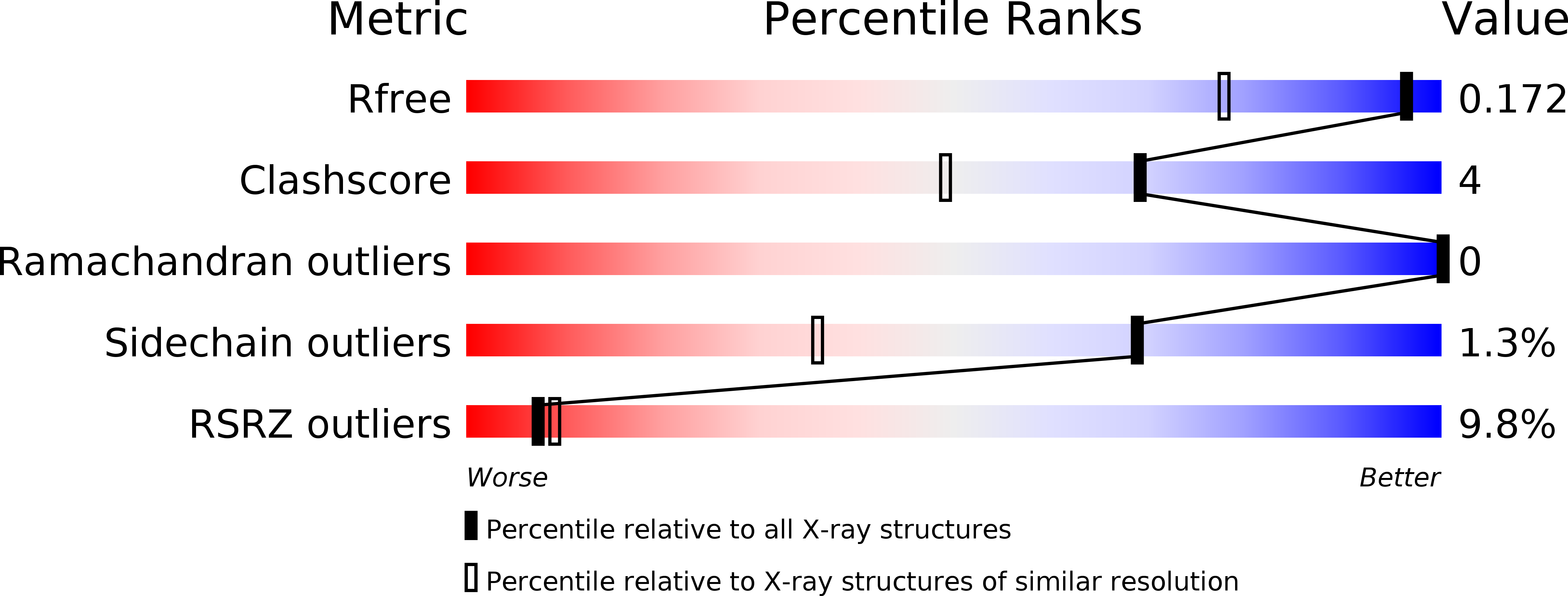
Resolution:
1.35 Å
R-Value Free:
0.15
R-Value Work:
0.13
R-Value Observed:
0.13
Space Group:
C 2 2 21
Macromolecular Entities
Polymer Type:polypeptide(L)
Molecule:plu4264
Gene (Uniprot):plu4264
Chain IDs:A, B
Chain Length:128
Number of Molecules:2
Biological Source:Photorhabdus luminescens subsp. laumondii
Modified Residue
Compound ID Chain ID Parent Comp ID Details 2D Image
MSE A MET SELENOMETHIONINE
Primary Citation
Structure of a cupin protein Plu4264 from Photorhabdus luminescens subsp. laumondii TTO1 at 1.35 angstrom resolution.
Proteins 83 383 388 (2015)
PMID: 25354690 DOI: 10.1002/prot.24705

Abstact

Proteins belonging to the cupin superfamily have a wide range of catalytic and noncatalytic functions. Cupin proteins commonly have the capacity to bind a metal ion with the metal frequently determining the function of the protein. We have been investigating the function of homologous cupin proteins that are conserved in more than 40 species of bacteria. To gain insights into the potential function of these proteins we have solved the structure of Plu4264 from Photorhabdus luminescens TTO1 at a resolution of 1.35 Å and identified manganese as the likely natural metal ligand of the protein.

Legend

Protein

Chemical

Disease

Primary Citation of related structures