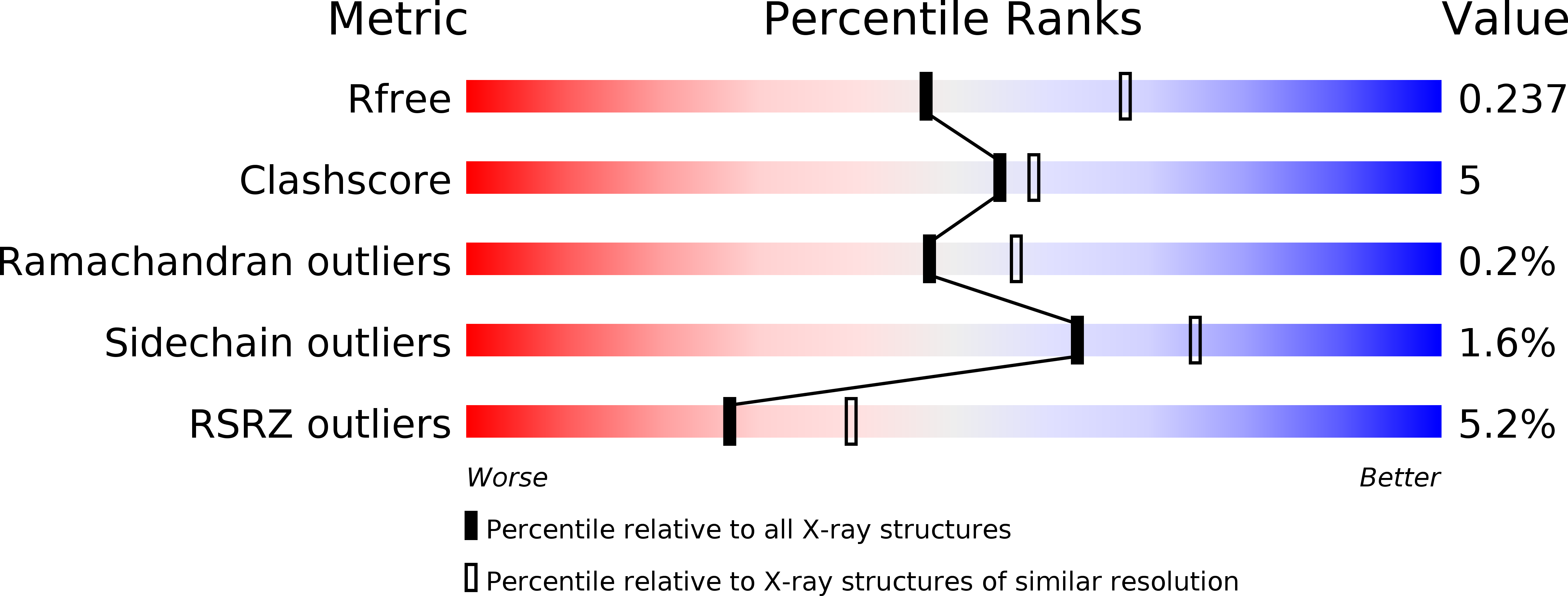
4MQE image
Entry Detail
PDB ID:
4MQE
Title:
Crystal structure of the extracellular domain of human GABA(B) receptor in the apo form
Biological Source:
Source Organism:
Host Organism:
PDB Version:
Deposition Date:
2013-09-16
Release Date:
2013-12-11
Method Details:
Experimental Method:
Resolution:
2.35 Å
R-Value Free:
0.23
R-Value Work:
0.20
R-Value Observed:
0.20
Space Group:
P 1 21 1
Macromolecular Entities
Polymer Type:polypeptide(L)
Description:Gamma-aminobutyric acid type B receptor subunit 1
Chain IDs:A
Chain Length:420
Number of Molecules:1
Biological Source:Homo sapiens
Polymer Type:polypeptide(L)
Description:Gamma-aminobutyric acid type B receptor subunit 2
Chain IDs:B
Chain Length:433
Number of Molecules:1
Biological Source:Homo sapiens
Modified Residue
Compound ID Chain ID Parent Comp ID Details 2D Image
ASN B ASN GLYCOSYLATION SITE
Ligand Molecules
Primary Citation
Structural mechanism of ligand activation in human GABA(B) receptor.
Nature 504 254 259 (2013)
PMID: 24305054 DOI: 10.1038/nature12725

Abstact

Human GABA(B) (γ-aminobutyric acid class B) receptor is a G-protein-coupled receptor central to inhibitory neurotransmission in the brain. It functions as an obligatory heterodimer of the subunits GBR1 and GBR2. Here we present the crystal structures of a heterodimeric complex between the extracellular domains of GBR1 and GBR2 in the apo, agonist-bound and antagonist-bound forms. The apo and antagonist-bound structures represent the resting state of the receptor; the agonist-bound complex corresponds to the active state. Both subunits adopt an open conformation at rest, and only GBR1 closes on agonist-induced receptor activation. The agonists and antagonists are anchored in the interdomain crevice of GBR1 by an overlapping set of residues. An antagonist confines GBR1 to the open conformation of the inactive state, whereas an agonist induces its domain closure for activation. Our data reveal a unique activation mechanism for GABA(B) receptor that involves the formation of a novel heterodimer interface between subunits.

Legend

Protein

Chemical

Disease

Primary Citation of related structures