4MPS image
Deposition Date 2013-09-13
Release Date 2013-09-25
Last Version Date 2024-11-27
Entry Detail
PDB ID:
4MPS
Keywords:
Title:
Crystal structure of rat Beta-galactoside alpha-2,6-sialyltransferase 1 (ST6GAL1), Northeast Structural Genomics Consortium Target RnR367A
Biological Source:
Source Organism:
Host Organism:
Method Details:
Experimental Method:
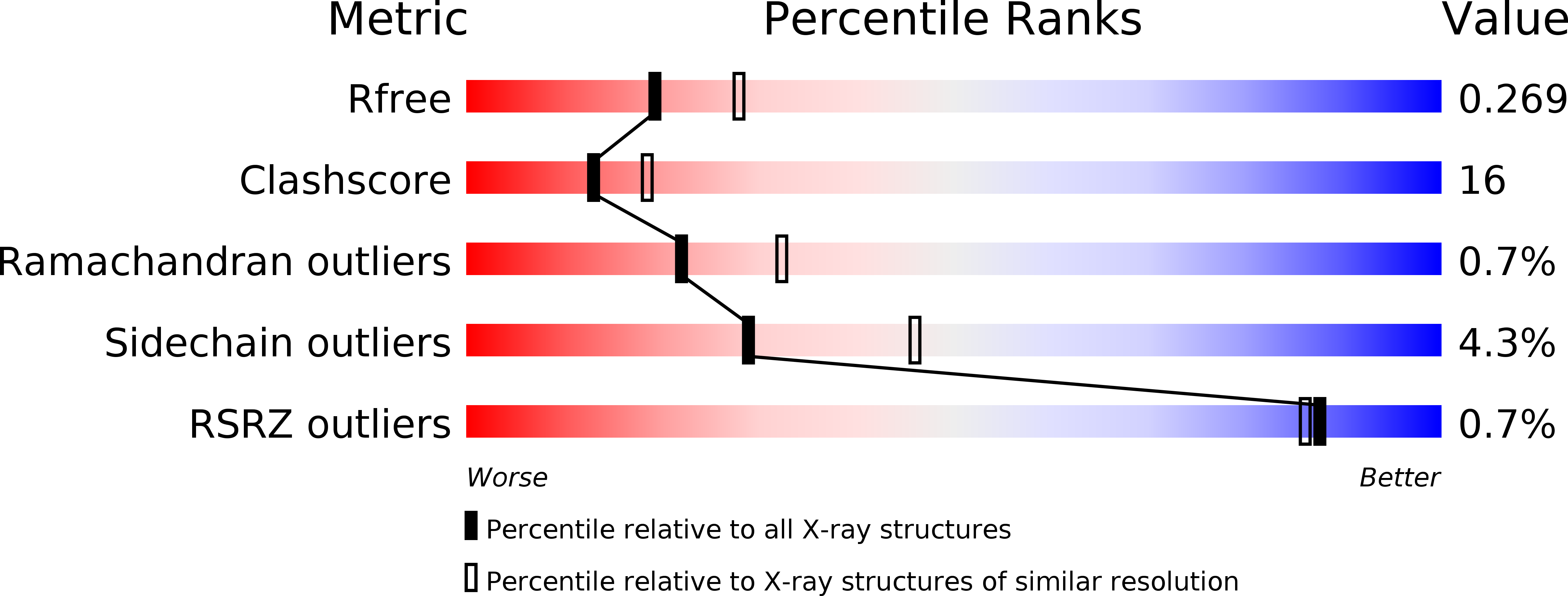
Resolution:
2.40 Å
R-Value Free:
0.25
R-Value Work:
0.20
R-Value Observed:
0.20
Space Group:
C 1 2 1
Macromolecular Entities
Polymer Type:polypeptide(L)
Molecule:Beta-galactoside alpha-2,6-sialyltransferase 1
Chain IDs:A, B
Chain Length:310
Number of Molecules:2
Biological Source:Rattus norvegicus
Modified Residue
Compound ID Chain ID Parent Comp ID Details 2D Image
ASN B ASN GLYCOSYLATION SITE
MSE A MET SELENOMETHIONINE
Ligand Molecules
Primary Citation
Enzymatic Basis for N-Glycan Sialylation: STRUCTURE OF RAT alpha 2,6-SIALYLTRANSFERASE (ST6GAL1) REVEALS CONSERVED AND UNIQUE FEATURES FOR GLYCAN SIALYLATION.
J.Biol.Chem. 288 34680 34698 (2013)
PMID: 24155237 DOI: 10.1074/jbc.M113.519041

Abstact

Glycan structures on glycoproteins and glycolipids play critical roles in biological recognition, targeting, and modulation of functions in animal systems. Many classes of glycan structures are capped with terminal sialic acid residues, which contribute to biological functions by either forming or masking glycan recognition sites on the cell surface or secreted glycoconjugates. Sialylated glycans are synthesized in mammals by a single conserved family of sialyltransferases that have diverse linkage and acceptor specificities. We examined the enzymatic basis for glycan sialylation in animal systems by determining the crystal structures of rat ST6GAL1, an enzyme that creates terminal α2,6-sialic acid linkages on complex-type N-glycans, at 2.4 Å resolution. Crystals were obtained from enzyme preparations generated in mammalian cells. The resulting structure revealed an overall protein fold broadly resembling the previously determined structure of pig ST3GAL1, including a CMP-sialic acid-binding site assembled from conserved sialylmotif sequence elements. Significant differences in structure and disulfide bonding patterns were found outside the sialylmotif sequences, including differences in residues predicted to interact with the glycan acceptor. Computational substrate docking and molecular dynamics simulations were performed to predict and evaluate the CMP-sialic acid donor and glycan acceptor interactions, and the results were compared with kinetic analysis of active site mutants. Comparisons of the structure with pig ST3GAL1 and a bacterial sialyltransferase revealed a similar positioning of donor, acceptor, and catalytic residues that provide a common structural framework for catalysis by the mammalian and bacterial sialyltransferases.

Legend

Protein

Chemical

Disease

Primary Citation of related structures