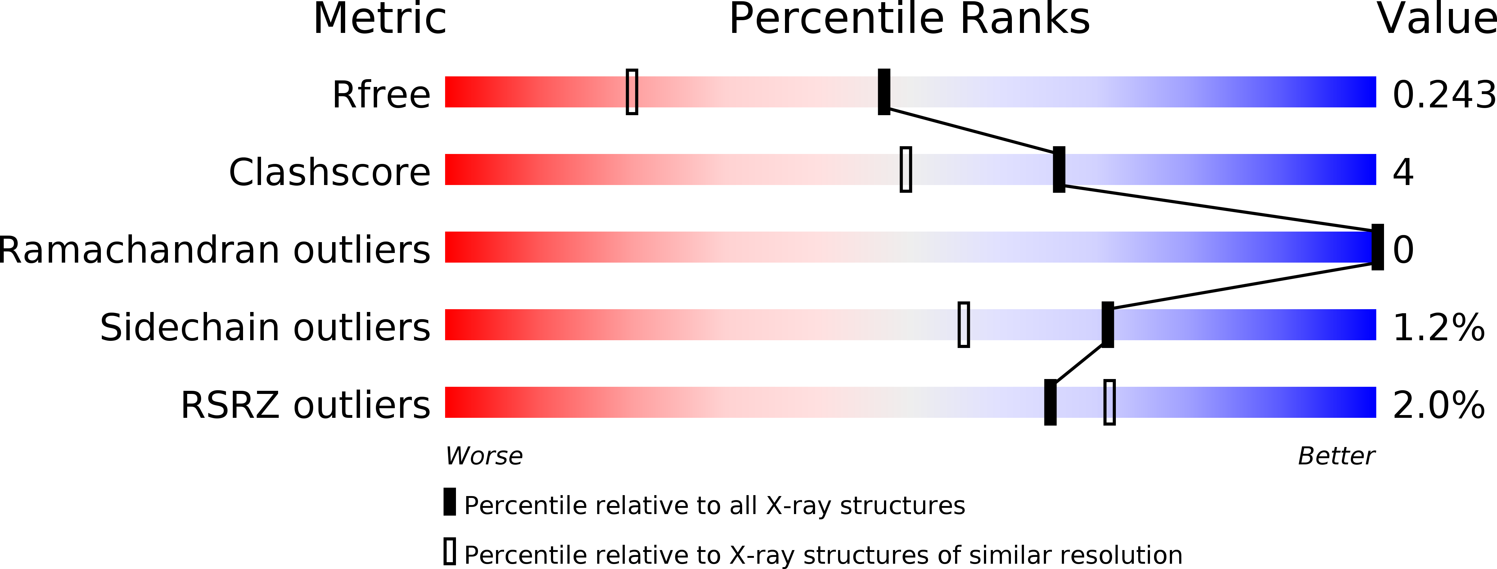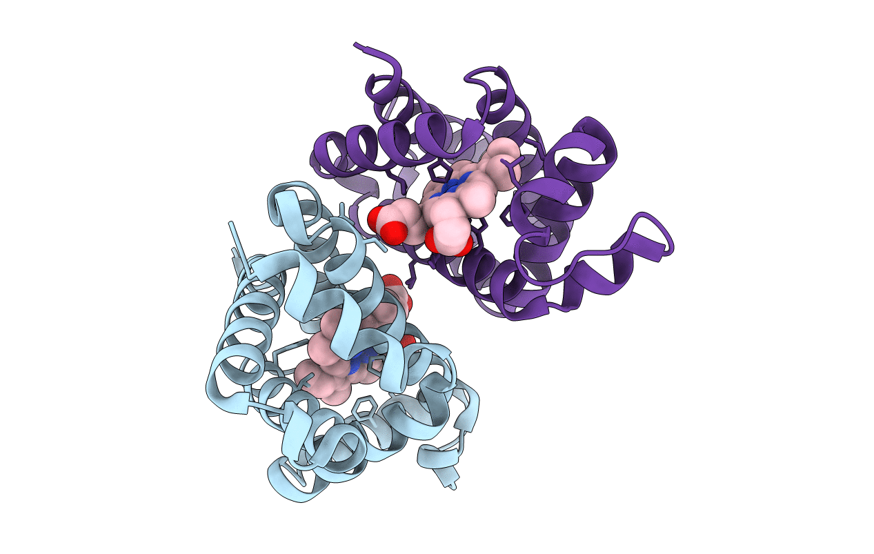
4MPM image
Deposition Date 2013-09-13
Release Date 2014-01-15
Last Version Date 2024-11-06
Entry Detail
PDB ID:
4MPM
Keywords:
Title:
Wild-type human neuroglobin
Biological Source:
Source Organism:
Homo sapiens (Taxon ID: 9606)
Host Organism:
Method Details:
Experimental Method:
Resolution:
1.74 Å
R-Value Free:
0.22
R-Value Work:
0.18
R-Value Observed:
0.18
Space Group:
P 21 21 2
Macromolecular Entities
Structures with similar UniProt ID
Protein Blast
Polymer Type:polypeptide(L)
Molecule:Neuroglobin
Gene (Uniprot):NGB
Chain IDs:A, B
Chain Length:151
Number of Molecules:2
Biological Source:Homo sapiens
Ligand Molecules
Primary Citation
The crystal structure of wild-type human brain neuroglobin reveals flexibility of the disulfide bond that regulates oxygen affinity.
Acta Crystallogr.,Sect.D 70 1005 1014 (2014)
PMID: 24699645 DOI: 10.1107/S1399004714000078

Abstact

Neuroglobin plays an important function in the supply of oxygen in nervous tissues. In human neuroglobin, a cysteine at position 46 in the loop connecting the C and D helices of the globin fold is presumed to form an intramolecular disulfide bond with Cys55. Rupture of this disulfide bridge stabilizes bi-histidyl haem hexacoordination, causing an overall decrease in the affinity for oxygen. Here, the first X-ray structure of wild-type human neuroglobin is reported at 1.74 Å resolution. This structure provides a direct observation of two distinct conformations of the CD region containing the intramolecular disulfide link and highlights internal cavities that could be involved in ligand migration and/or are necessary to enable the conformational transition between the low and high oxygen-affinity states following S-S bond formation.

Legend

Protein

Chemical

Disease

Primary Citation of related structures