
Deposition Date
2013-09-06
Release Date
2014-04-02
Last Version Date
2025-03-26
Entry Detail
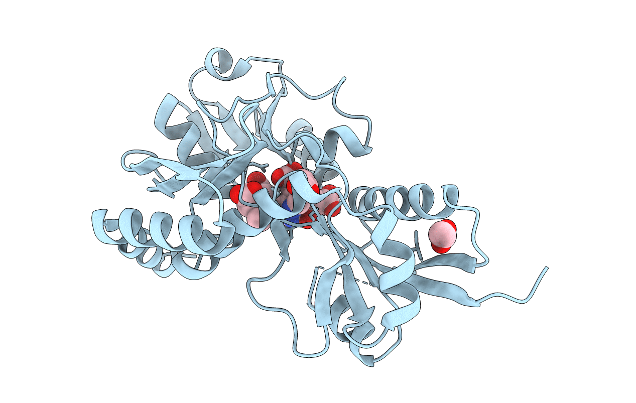
PDB ID:
4MLQ
Keywords:
Title:
Crystal structure of Bacillus megaterium porphobilinogen deaminase
Biological Source:
Source Organism(s):
Bacillus megaterium (Taxon ID: 1404)
Expression System(s):
Method Details:
Experimental Method:
Resolution:
1.60 Å
R-Value Free:
0.24
R-Value Work:
0.16
R-Value Observed:
0.16
Space Group:
P 21 21 21
