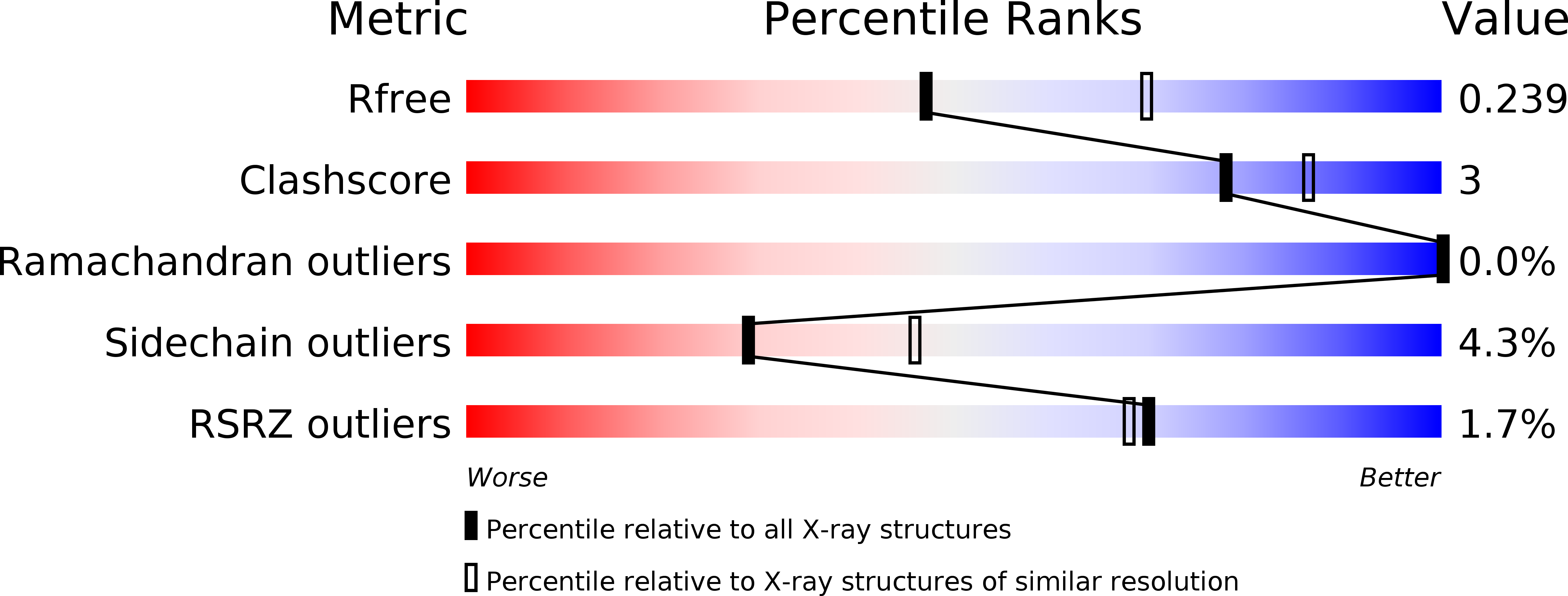
4MJO image
Deposition Date 2013-09-04
Release Date 2013-11-06
Last Version Date 2023-09-20
Entry Detail
PDB ID:
4MJO
Title:
Human liver fructose-1,6-bisphosphatase(d-fructose-1,6-bisphosphate, 1-phosphohydrolase) (e.c.3.1.3.11) complexed with the allosteric inhibitor 3
Biological Source:
Source Organism:
Homo sapiens (Taxon ID: 9606)
Method Details:
Experimental Method:
Resolution:
2.40 Å
R-Value Free:
0.23
R-Value Work:
0.20
R-Value Observed:
0.20
Space Group:
P 1 21 1
Macromolecular Entities
Polymer Type:polypeptide(L)
Molecule:Fructose-1,6-bisphosphatase 1
Gene (Uniprot):FBP1
Chain IDs:A, B, C, D, E, F, G, H
Chain Length:338
Number of Molecules:8
Biological Source:Homo sapiens
Ligand Molecules
Primary Citation
Determination of protein-ligand binding constants of a cooperatively regulated tetrameric enzyme using electrospray mass spectrometry.
Acs Chem.Biol. 9 218 226 (2014)
PMID: 24128068 DOI: 10.1021/cb4007002

Abstact

This study highlights the benefits of nano electrospray ionization mass spectrometry (nanoESI-MS) as a fast and label-free method not only for determination of dissociation constants (KD) of a cooperatively regulated enzyme but also to better understand the mechanism of enzymatic cooperativity of multimeric proteins. We present an approach to investigate the allosteric mechanism in the binding of inhibitors to the homotetrameric enzyme fructose 1,6-bisphosphatase (FBPase), a potential therapeutic target for glucose control in type 2 diabetes. A series of inhibitors binding at an allosteric site of FBPase were investigated to determine their KDs by nanoESI-MS. The KDs determined by ESI-MS correlate very well with IC50 values in solution. The Hill coefficients derived from nanoESI-MS suggest positive cooperativity. From single-point measurements we could obtain information on relative potency, stoichiometry, conformational changes, and mechanism of cooperativity. A new X-ray crystal structure of FBPase tetramer binding ligand 3 in a 4:4 stoichiometry is also reported. NanoESI-MS-based results match the current understanding of the investigated system and are in agreement with the X-ray structural data, but provide additional mechanistic insight on the ligand binding, due to the better dynamic resolution. This method offers a powerful approach for studying other proteins with allosteric binding sites, as well.

Legend

Protein

Chemical

Disease

Primary Citation of related structures