Deposition Date
2013-08-28
Release Date
2014-08-13
Last Version Date
2024-02-28
Entry Detail
PDB ID:
4MGJ
Keywords:
Title:
Crystal structure of cytochrome P450 2B4 F429H in complex with 4-CPI
Biological Source:
Source Organism(s):
Oryctolagus cuniculus (Taxon ID: 9986)
Expression System(s):
Method Details:
Experimental Method:
Resolution:
2.41 Å
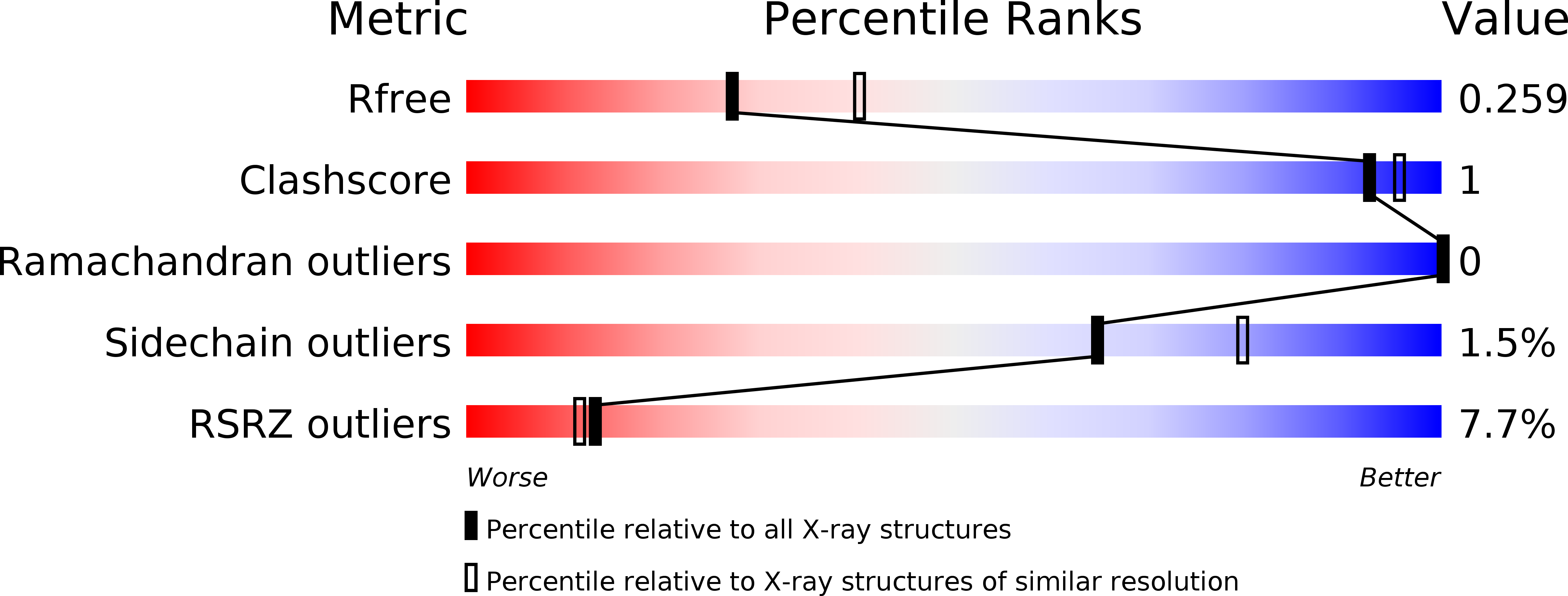
R-Value Free:
0.24
R-Value Work:
0.21
R-Value Observed:
0.22
Space Group:
P 31 2 1