4MEC image
Deposition Date 2013-08-26
Release Date 2014-08-27
Last Version Date 2023-11-08
Entry Detail
PDB ID:
4MEC
Keywords:
Title:
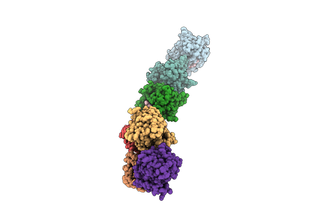
Crystal structure of RAT Heme oxygenase-1 in complex with ZN(II)-Protoporphyrin IX
Biological Source:
Source Organism:
Host Organism:
Method Details:
Experimental Method:
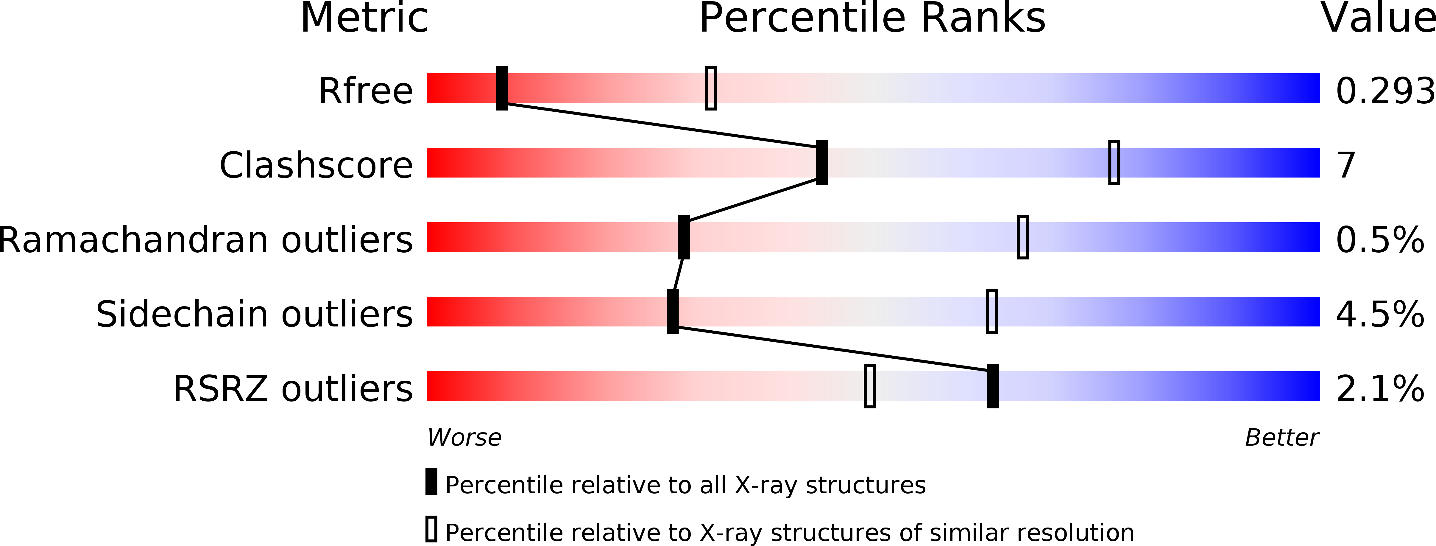
Resolution:
3.20 Å
R-Value Free:
0.29
R-Value Work:
0.24
R-Value Observed:
0.25
Space Group:
P 1
Macromolecular Entities
Polymer Type:polypeptide(L)
Molecule:Heme oxygenase 1
Gene (Uniprot):Hmox1
Chain IDs:A, B, C, D, E, F, G
Chain Length:232
Number of Molecules:7
Biological Source:Rattus norvegicus
Ligand Molecules
Primary Citation
Distal regulation of heme binding of heme oxygenase-1 mediated by conformational fluctuations
Biochemistry 54 340 348 (2015)
PMID: 25496210 DOI: 10.1021/bi5009694

Abstact

Heme oxygenase-1 (HO-1) is an enzyme that catalyzes the oxidative degradation of heme. Since free heme is toxic to cells, rapid degradation of heme is important for maintaining cellular health. There have been useful mechanistic studies of the HO reaction based on crystal structures; however, how HO-1 recognizes heme is not completely understood because the crystal structure of heme-free rat HO-1 lacks electron densities for A-helix that ligates heme. In this study, we characterized conformational dynamics of HO-1 using NMR to elucidate the mechanism by which HO-1 recognizes heme. NMR relaxation experiments showed that the heme-binding site in heme-free HO-1 fluctuates in concert with a surface-exposed loop and transiently forms a partially unfolded structure. Because the fluctuating loop is located over 17 Å distal from the heme-binding site and its conformation is nearly identical among different crystal structures including catalytic intermediate states, the function of the loop has been unexamined. In the course of elucidating its function, we found interesting mutations in this loop that altered activity but caused little change to the conformation. The Phe79Ala mutation in the loop changed the conformational dynamics of the heme-binding site. Furthermore, the heme binding kinetics of the mutant was slower than that of the wild type. Hence, we concluded that the distal loop is involved in the regulation of the conformational change for heme binding through the conformational fluctuations. Similar to other enzymes, HO-1 effectively promotes its function using the identified distal sites, which might be potential targets for protein engineering.

Legend

Protein

Chemical

Disease

Primary Citation of related structures