4ME7 image
Deposition Date 2013-08-25
Release Date 2013-10-23
Last Version Date 2024-11-20
Entry Detail
PDB ID:
4ME7
Title:
Crystal structure of Bacillus subtilis toxin MazF in complex with cognate antitoxin MazE
Biological Source:
Source Organism:
Host Organism:
Method Details:
Experimental Method:
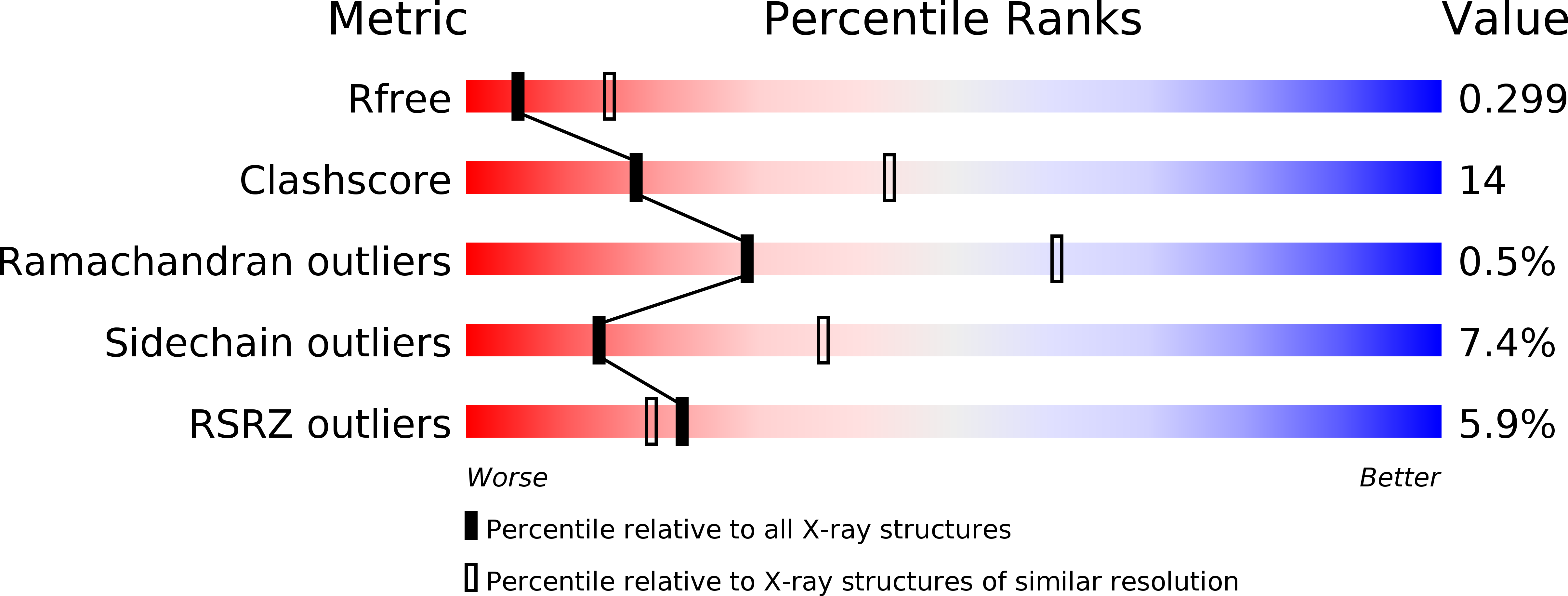
Resolution:
2.92 Å
R-Value Free:
0.29
R-Value Work:
0.25
R-Value Observed:
0.25
Space Group:
P 21 21 21
Macromolecular Entities
Structures with similar UniProt ID
Protein Blast
Polymer Type:polypeptide(L)
Molecule:mRNA interferase EndoA
Gene (Uniprot):ndoA
Chain IDs:A, B, C, D
Chain Length:116
Number of Molecules:4
Biological Source:Bacillus subtilis subsp. subtilis
Structures with similar UniProt ID
Protein Blast
Polymer Type:polypeptide(L)
Molecule:Antitoxin EndoAI
Gene (Uniprot):ndoAI
Chain IDs:E, F
Chain Length:94
Number of Molecules:2
Biological Source:Bacillus subtilis subsp. subtilis
Modified Residue
Compound ID Chain ID Parent Comp ID Details 2D Image
MSE A MET SELENOMETHIONINE
Primary Citation
Structural Basis of mRNA Recognition and Cleavage by Toxin MazF and Its Regulation by Antitoxin MazE in Bacillus subtilis.
Mol.Cell 52 447 458 (2013)
PMID: 24120662 DOI: 10.1016/j.molcel.2013.09.006

Abstact

MazF is an mRNA interferase, which, upon activation during stress conditions, cleaves mRNAs in a sequence-specific manner, resulting in cellular growth arrest. During normal growth conditions, the MazF toxin is inactivated through binding to its cognate antitoxin, MazE. How MazF specifically recognizes its mRNA target and carries out cleavage and how the formation of the MazE-MazF complex inactivates MazF remain unclear. We present crystal structures of MazF in complex with mRNA substrate and antitoxin MazE in Bacillus subtilis. The structure of MazF in complex with uncleavable UUdUACAUAA RNA substrate defines the molecular basis underlying the sequence-specific recognition of UACAU and the role of residues involved in the cleavage through site-specific mutational studies. The structure of the heterohexameric (MazF)2-(MazE)2-(MazF)2 complex in Bacillus subtilis, supplemented by mutational data, demonstrates that the positioning of the C-terminal helical segment of MazE within the RNA-binding channel of the MazF dimer prevents mRNA binding and cleavage by MazF.

Legend

Protein

Chemical

Disease

Primary Citation of related structures