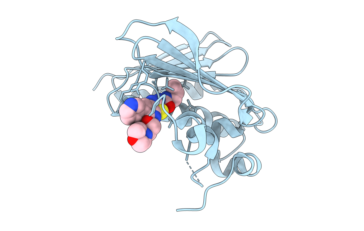
4MBC image
Entry Detail
PDB ID:
4MBC
Title:
Structure of Streptococcus pneumonia ParE in complex with AZ13053807
Biological Source:
Source Organism:
Host Organism:
PDB Version:
Deposition Date:
2013-08-19
Release Date:
2013-10-16
Method Details:
Experimental Method:
Resolution:
1.75 Å
R-Value Free:
0.22
R-Value Work:
0.18
R-Value Observed:
0.18
Space Group:
C 2 2 21
Macromolecular Entities
Polymer Type:polypeptide(L)
Description:DNA topoisomerase IV, B subunit
Chain IDs:A
Chain Length:226
Number of Molecules:1
Biological Source:Streptococcus pneumoniae
Ligand Molecules
Primary Citation

Abstact

A pharmacophore-based search led to the identification of thiazolopyridine ureas as a novel scaffold with antitubercular activity acting through inhibition of DNA Gyrase B (GyrB) ATPase. Evaluation of the binding mode of thiazolopyridines in a Mycobacterium tuberculosis (Mtb) GyrB homology model prompted exploration of the side chains at the thiazolopyridine ring C-5 position to access the ribose/solvent pocket. Potent compounds with GyrB IC50 ≤ 1 nM and Mtb MIC ≤ 0.1 μM were obtained with certain combinations of side chains at the C-5 position and heterocycles at the C-6 position of the thiazolopyridine core. Substitutions at C-5 also enabled optimization of the physicochemical properties. Representative compounds were cocrystallized with Streptococcus pneumoniae (Spn) ParE; these confirmed the binding modes predicted by the homology model. The target link to GyrB was confirmed by genetic mapping of the mutations conferring resistance to thiazolopyridine ureas. The compounds are bactericidal in vitro and efficacious in vivo in an acute murine model of tuberculosis.

Legend

Protein

Chemical

Disease

Primary Citation of related structures