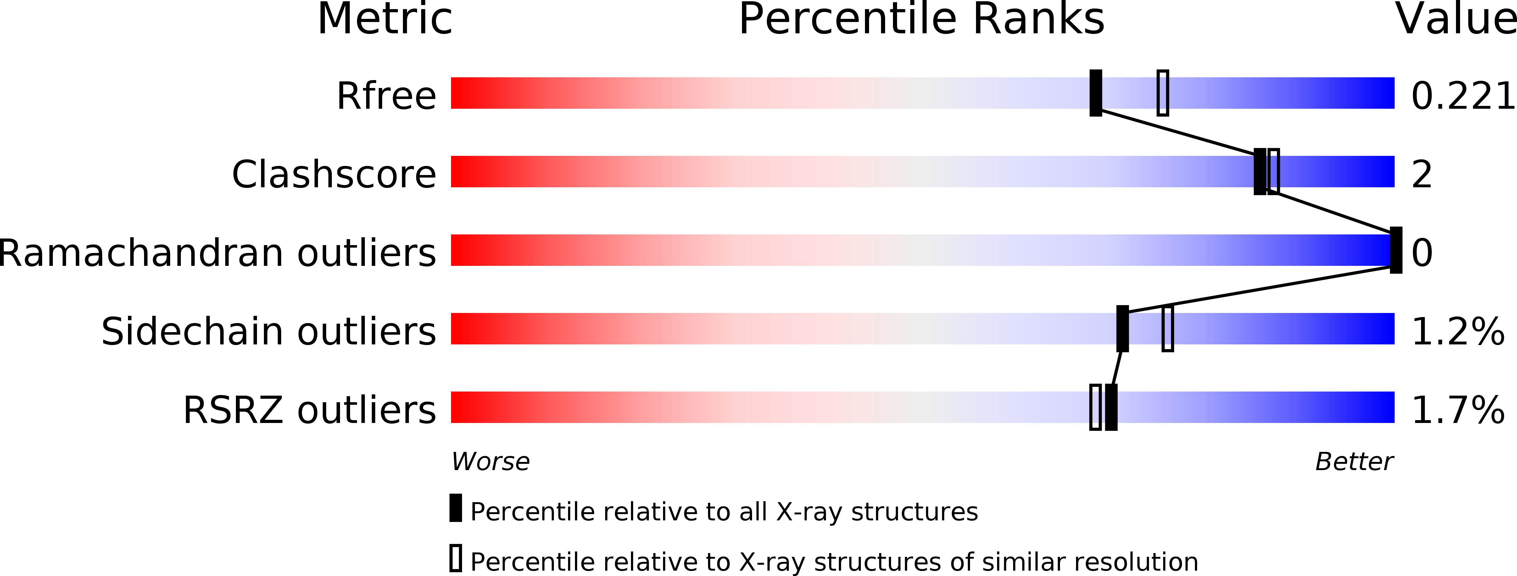
4M9G image
Deposition Date 2013-08-14
Release Date 2013-10-16
Last Version Date 2024-02-28
Entry Detail
PDB ID:
4M9G
Keywords:
Title:
DNA Polymerase Beta E295K Binary Complex
Biological Source:
Source Organism:
Homo sapiens (Taxon ID: 9606)
Host Organism:
Method Details:
Experimental Method:
Resolution:
2.01 Å
R-Value Free:
0.22
R-Value Work:
0.18
R-Value Observed:
0.18
Space Group:
P 1 21 1
Macromolecular Entities
Structures with similar UniProt ID
Protein Blast
Polymer Type:polypeptide(L)
Molecule:DNA polymerase beta
Gene (Uniprot):POLB
Mutagens:E295K
Chain IDs:A
Chain Length:335
Number of Molecules:1
Biological Source:Homo sapiens
Polymer Type:polydeoxyribonucleotide
Molecule:DNA Downstream Strand
Chain IDs:D
Chain Length:5
Number of Molecules:1
Biological Source:
Polymer Type:polydeoxyribonucleotide
Molecule:DNA Primer Strand
Chain IDs:C (auth: P)
Chain Length:10
Number of Molecules:1
Biological Source:
Polymer Type:polydeoxyribonucleotide
Molecule:DNA Template Strand
Chain IDs:B (auth: T)
Chain Length:16
Number of Molecules:1
Biological Source:
Primary Citation
The E295K Cancer Variant of Human Polymerase beta Favors the Mismatch Conformational Pathway during Nucleotide Selection.
J.Biol.Chem. 288 34850 34860 (2013)
PMID: 24133209 DOI: 10.1074/jbc.M113.510891

Abstact

DNA polymerase β (pol β) is responsible for gap filling synthesis during repair of damaged DNA as part of the base excision repair pathway. Human pol β mutations were recently identified in a high percentage (∼30%) of tumors. Characterization of specific cancer variants is particularly useful to further the understanding of the general mechanism of pol β while providing context to disease contribution. We showed that expression of the carcinoma variant E295K induces cellular transformation. The poor polymerase activity exhibited by the variant was hypothesized to be caused by the destabilization of proper active site assembly by the glutamate to lysine mutation. Here, we show that this variant exhibits an unusual preference for binding dCTP opposite a templating adenine over the cognate dTTP. Biochemical studies indicate that the noncognate competes with the cognate nucleotide for binding to the polymerase active site with the noncognate incorporation a function of higher affinity and not increased activity. In the crystal structure of the variant bound to dA:dCTP, the fingers domain closes around the mismatched base pair. Nucleotide incorporation is hindered because key residues in the polymerase active site are not properly positioned for nucleotidyl transfer. In contrast to the noncognate dCTP, neither the cognate dTTP nor its nonhydrolyzable analog induced fingers closure, as isomorphous difference Fourier maps show that the cognate nucleotides are bound to the open state of the polymerase. Comparison with published structures provides insight into the structural rearrangements within pol β that occur during the process of nucleotide discrimination.

Legend

Protein

Chemical

Disease

Primary Citation of related structures