
Deposition Date
2013-08-14
Release Date
2014-03-12
Last Version Date
2024-02-28
Entry Detail
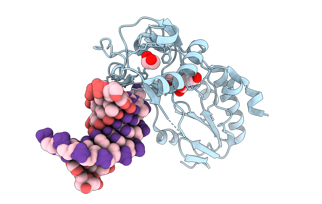
PDB ID:
4M94
Keywords:
Title:
d(ATCCGTTATAACGGAT) complexed with Moloney Murine Leukemia virus reverse transcriptase catalytic fragment
Biological Source:
Source Organism(s):
Moloney murine leukemia virus isolate Shinnick (Taxon ID: 928306)
synthetic construct (Taxon ID: 32630)
synthetic construct (Taxon ID: 32630)
Expression System(s):
Method Details:
Experimental Method:
Resolution:
2.14 Å
R-Value Free:
0.23
R-Value Work:
0.20
R-Value Observed:
0.20
Space Group:
P 21 21 2
