4M6J image
Deposition Date 2013-08-09
Release Date 2013-09-25
Last Version Date 2023-09-20
Entry Detail
PDB ID:
4M6J
Keywords:
Title:
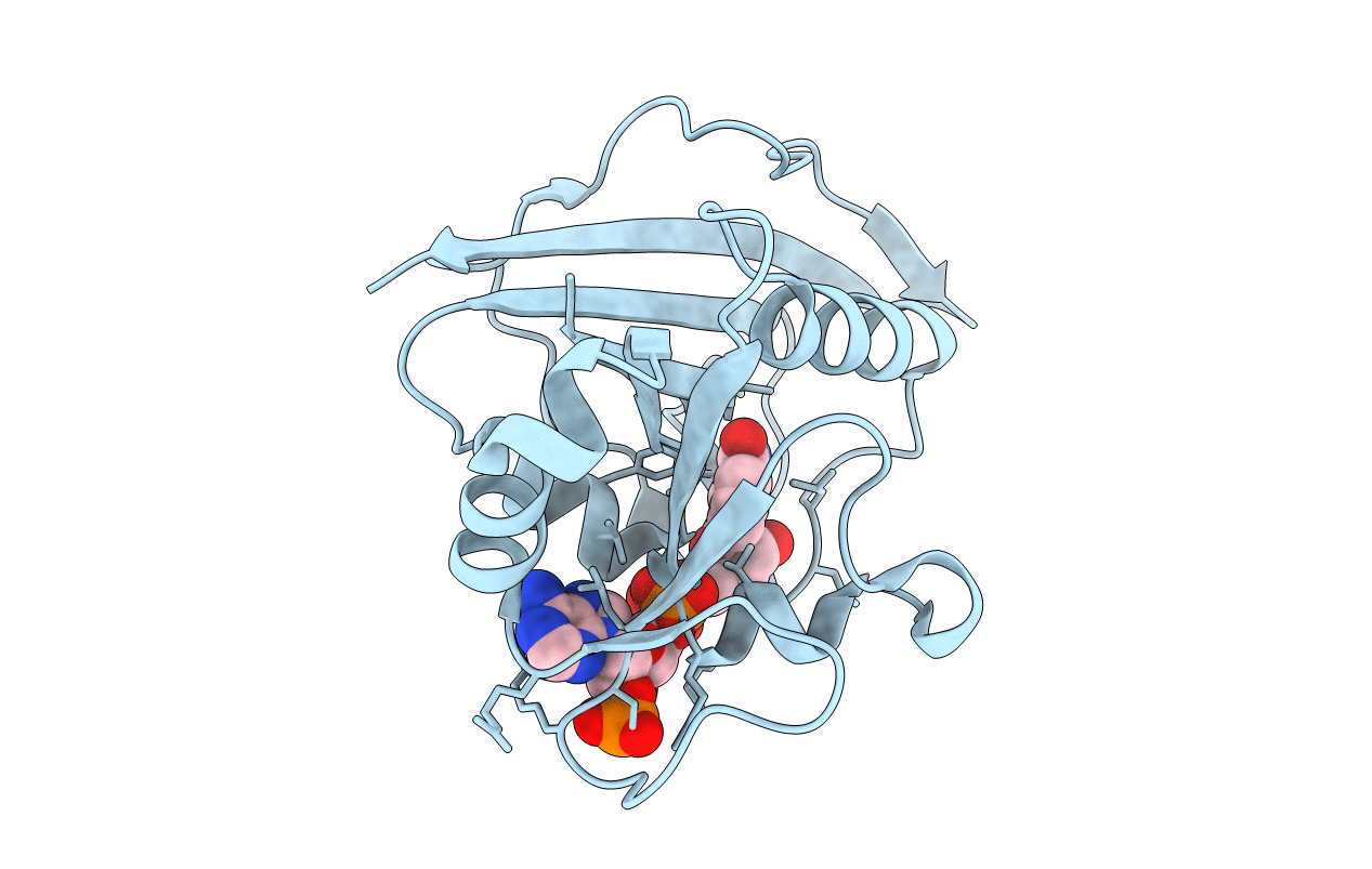
Crystal structure of human dihydrofolate reductase (DHFR) bound to NADPH
Biological Source:
Source Organism:
Homo sapiens (Taxon ID: 9606)
Host Organism:
Method Details:
Experimental Method:
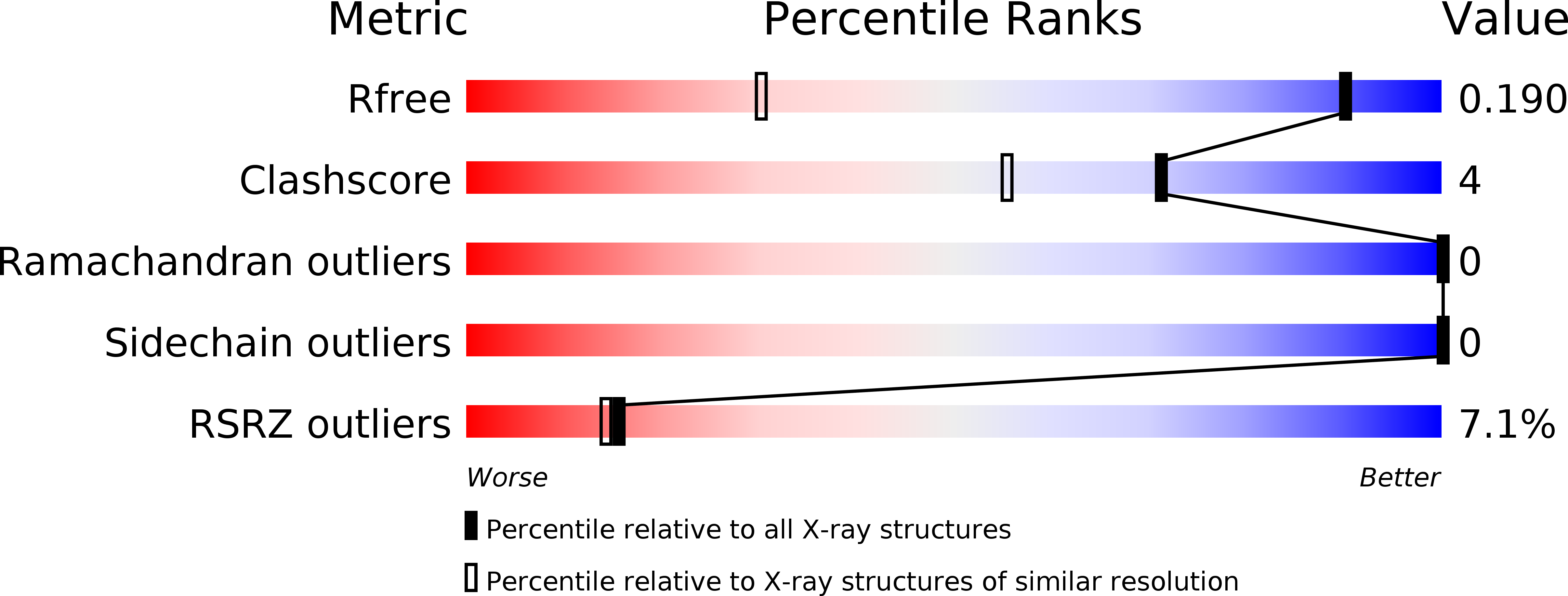
Resolution:
1.20 Å
R-Value Free:
0.18
R-Value Work:
0.16
R-Value Observed:
0.16
Space Group:
C 2 2 21
Macromolecular Entities
Polymer Type:polypeptide(L)
Molecule:Dihydrofolate reductase
Gene (Uniprot):DHFR
Chain IDs:A
Chain Length:187
Number of Molecules:1
Biological Source:Homo sapiens
Ligand Molecules
Primary Citation
Divergent evolution of protein conformational dynamics in dihydrofolate reductase.
Nat.Struct.Mol.Biol. 20 1243 1249 (2013)
PMID: 24077226 DOI: 10.1038/nsmb.2676

Abstact

Molecular evolution is driven by mutations, which may affect the fitness of an organism and are then subject to natural selection or genetic drift. Analysis of primary protein sequences and tertiary structures has yielded valuable insights into the evolution of protein function, but little is known about the evolution of functional mechanisms, protein dynamics and conformational plasticity essential for activity. We characterized the atomic-level motions across divergent members of the dihydrofolate reductase (DHFR) family. Despite structural similarity, Escherichia coli and human DHFRs use different dynamic mechanisms to perform the same function, and human DHFR cannot complement DHFR-deficient E. coli cells. Identification of the primary-sequence determinants of flexibility in DHFRs from several species allowed us to propose a likely scenario for the evolution of functionally important DHFR dynamics following a pattern of divergent evolution that is tuned by cellular environment.

Legend

Protein

Chemical

Disease

Primary Citation of related structures