Deposition Date
2013-07-31
Release Date
2014-01-08
Last Version Date
2024-10-30
Entry Detail
PDB ID:
4LZ4
Keywords:
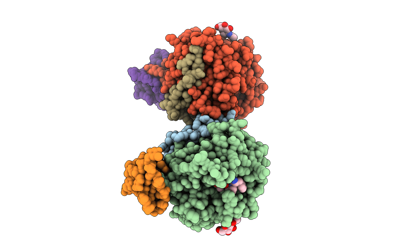
Title:
X-ray structure of the complex between human thrombin and the TBA deletion mutant lacking thymine 3 nucleobase
Biological Source:
Source Organism(s):
Homo sapiens (Taxon ID: 9606)
Method Details:
Experimental Method:
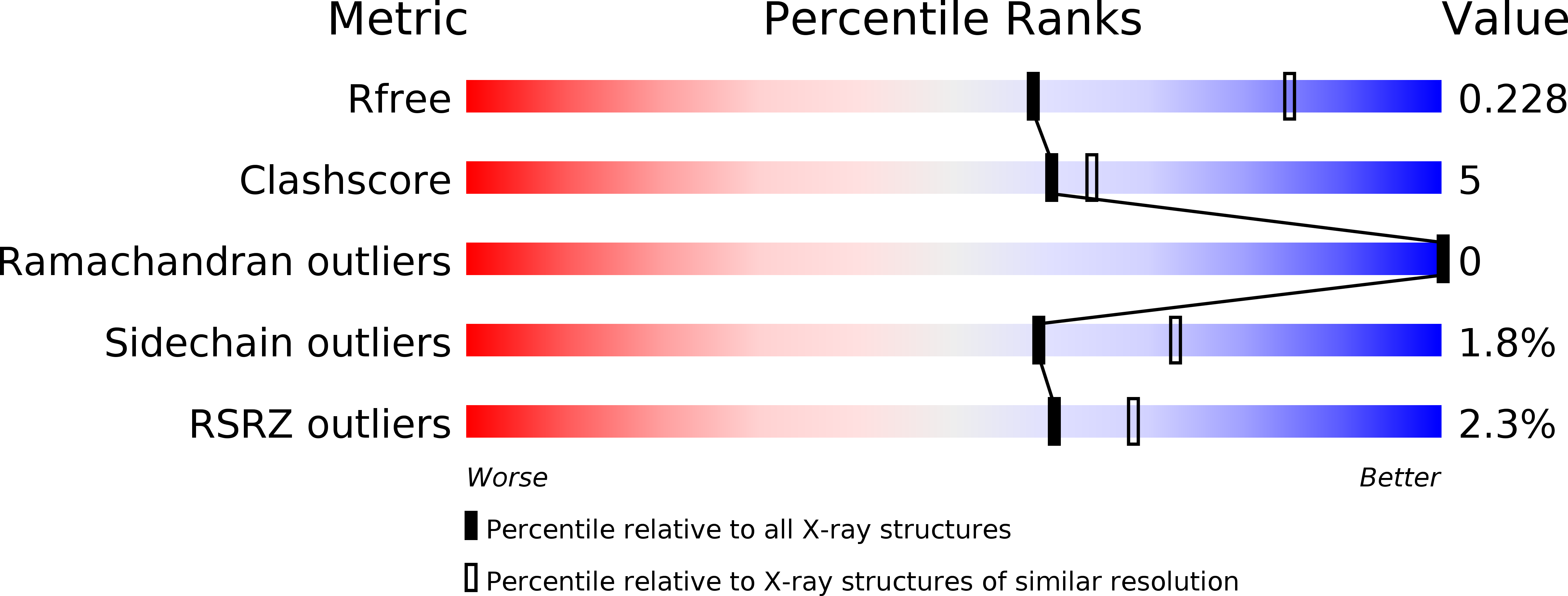
Resolution:
2.56 Å
R-Value Free:
0.22
R-Value Work:
0.16
R-Value Observed:
0.17
Space Group:
P 1 21 1