4LY4 image
Deposition Date 2013-07-30
Release Date 2013-08-28
Last Version Date 2023-09-20
Entry Detail
PDB ID:
4LY4
Keywords:
Title:
Crystal structure of peptidoglycan deacetylase (HP0310) with Zinc from Helicobacter pylori
Biological Source:
Source Organism:
Host Organism:
Method Details:
Experimental Method:
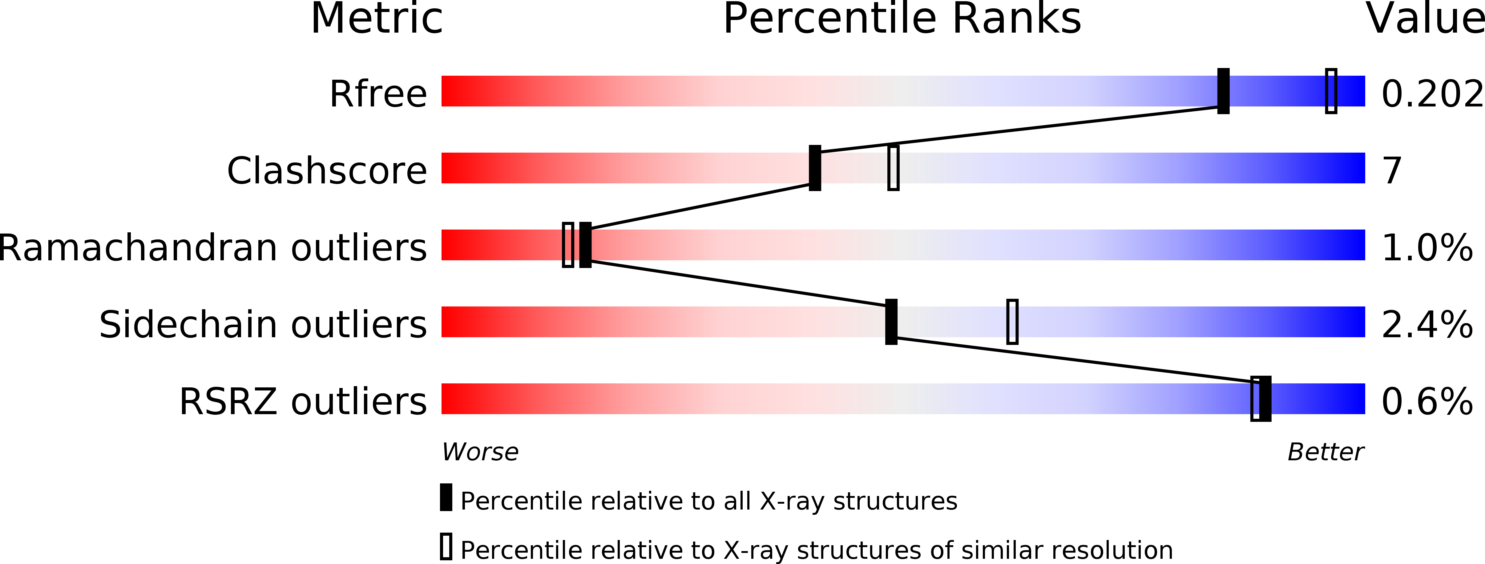
Resolution:
2.20 Å
R-Value Free:
0.20
R-Value Work:
0.16
R-Value Observed:
0.17
Space Group:
I 2 2 2
Macromolecular Entities
Polymer Type:polypeptide(L)
Molecule:peptidoglycan deacetylase
Gene (Uniprot):pgdA
Chain IDs:A, B, C, D
Chain Length:326
Number of Molecules:4
Biological Source:Helicobacter pylori
Ligand Molecules
Primary Citation
Characterization of the divalent metal binding site of bacterial polysaccharide deacetylase using crystallography and quantum chemical calculations.
Proteins 82 1311 1318 (2014)
PMID: 24346839 DOI: 10.1002/prot.24497

Abstact

Peptidoglycan deacetlyase (HP0310, HpPgdA) from the gram-negative pathogen Helicobacter pylori, is the enzyme responsible for a peptidoglycan modification that counteracts the host immune response. In a recent study, we determined the crystallographic structure of the enzyme, which is a homo-tetramer (Shaik et al., PloS One 2011;6:e19207). The metal-binding site, which is essential for the enzyme's catalytic activity, is visible within the structure, but we were unable to identify the nature of the metal itself. In this study, we have obtained a higher-resolution crystal structure of the enzyme, which shows that the ion bound is, in fact, zinc. Analysis of the structure of the four sites, one per monomer, and quantum chemical calculations of models of the site in the presence of different divalent metal ions show an intrinsic preference for zinc, but also significant flexibility of the site so that binding of other ions can eventually occur.

Legend

Protein

Chemical

Disease

Primary Citation of related structures