Deposition Date
2013-07-19
Release Date
2014-07-30
Last Version Date
2024-02-28
Entry Detail
PDB ID:
4LQS
Keywords:
Title:
Crystal structure of the Cbk1-Mob2 kinase-coactivator complex
Biological Source:
Source Organism(s):
Saccharomyces cerevisiae (Taxon ID: 559292)
Expression System(s):
Method Details:
Experimental Method:
Resolution:
3.30 Å
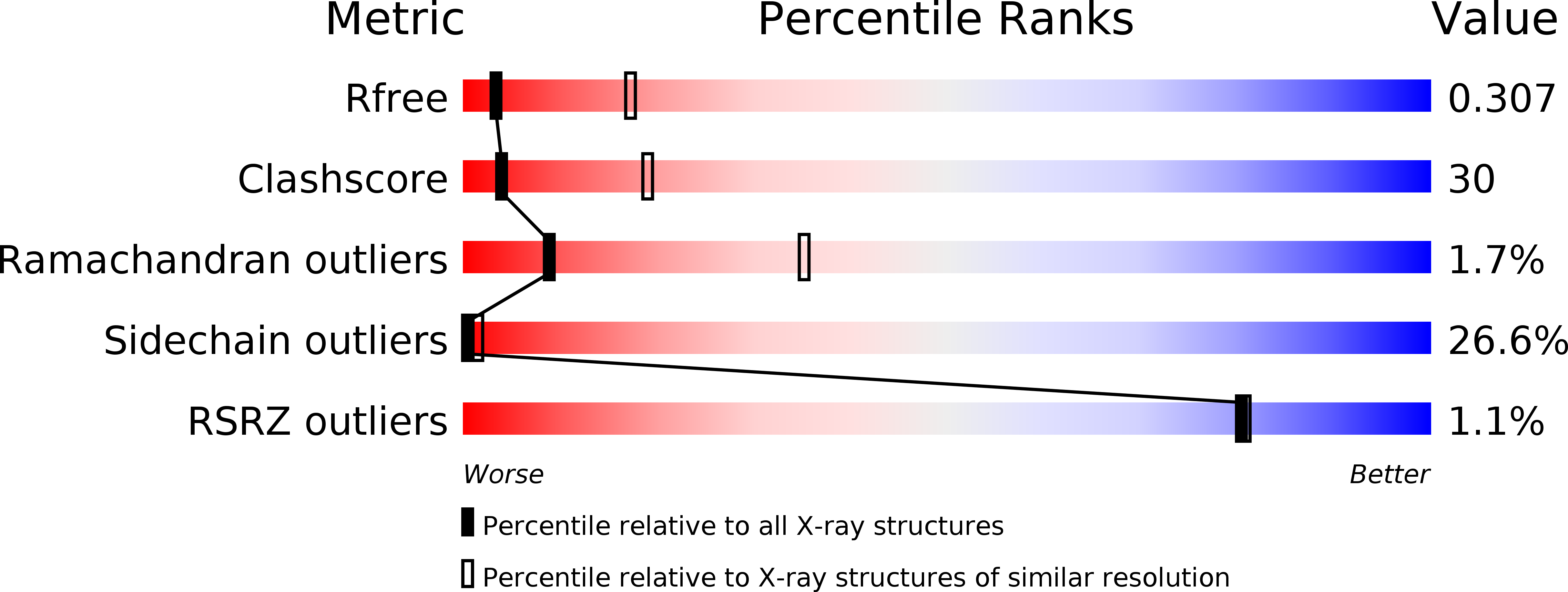
R-Value Free:
0.30
R-Value Work:
0.27
R-Value Observed:
0.27
Space Group:
C 1 2 1