4LP6 image
Deposition Date 2013-07-15
Release Date 2013-10-23
Last Version Date 2024-03-13
Entry Detail
PDB ID:
4LP6
Title:
Crystal Structure of Human Carbonic Anhydrase II in complex with a quinoline oligoamide foldamer
Biological Source:
Source Organism:
Homo sapiens (Taxon ID: 9606)
Host Organism:
Method Details:
Experimental Method:
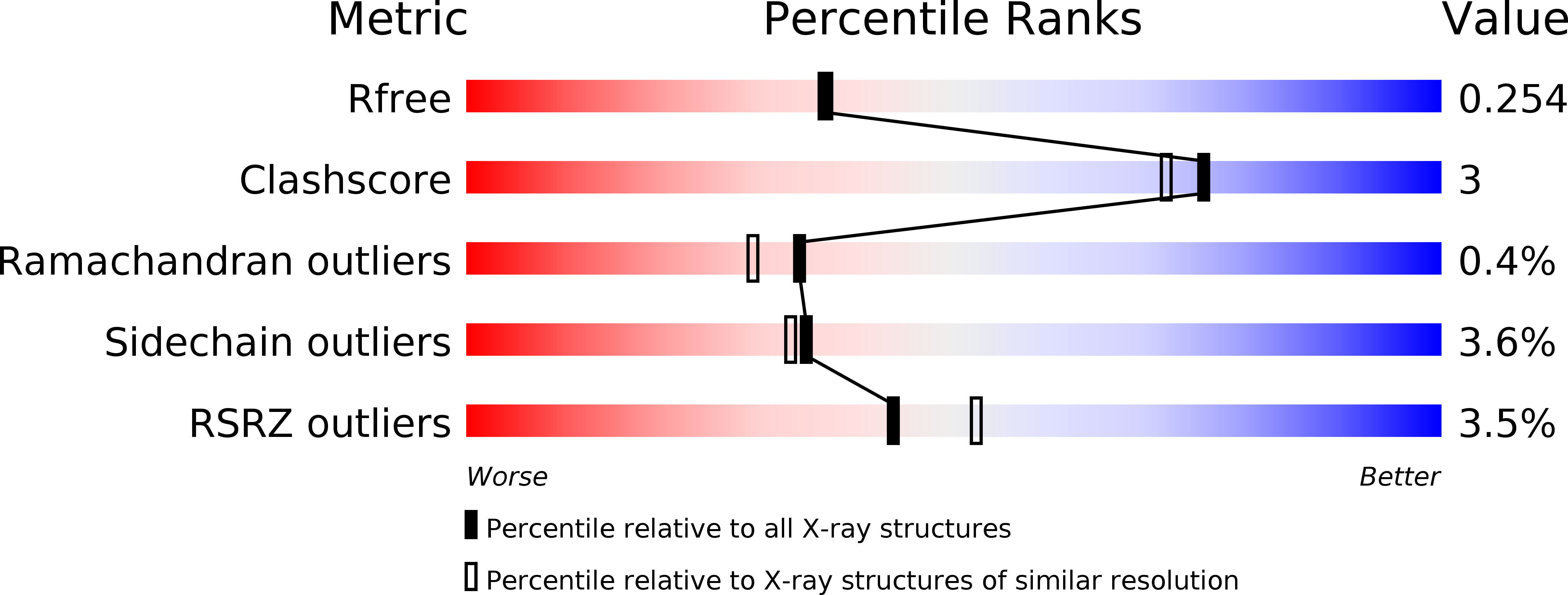
Resolution:
2.15 Å
R-Value Free:
0.25
R-Value Work:
0.19
R-Value Observed:
0.19
Space Group:
P 1 21 1
Macromolecular Entities
Polymer Type:polypeptide(L)
Molecule:Carbonic anhydrase 2
Gene (Uniprot):CA2
Chain IDs:A, B
Chain Length:260
Number of Molecules:2
Biological Source:Homo sapiens
Primary Citation
Structure of a complex formed by a protein and a helical aromatic oligoamide foldamer at 2.1 angstrom resolution.
Angew.Chem.Int.Ed.Engl. 53 883 887 (2014)
PMID: 24288253 DOI: 10.1002/anie.201309160

Abstact

In the search of molecules that could recognize sizeable areas of protein surfaces, a series of ten helical aromatic oligoamide foldamers was synthesized on solid phase. The foldamers comprise three to five monomers carrying various proteinogenic side chains, and exist as racemic mixtures of interconverting right-handed and left-handed helices. Functionalization of the foldamers by a nanomolar ligand of human carbonic anhydrase II (HCA) ensured that they would be held in close proximity to the protein surface. Foldamer-protein interactions were screened by circular dichroism (CD). One foldamer displayed intense CD bands indicating that a preferred helix handedness is induced upon interacting with the protein surface. The crystal structure of the complex between this foldamer and HCA could be resolved at 2.1 Å resolution and revealed a number of unanticipated protein-foldamer, foldamer-foldamer, and protein-protein interactions.

Legend

Protein

Chemical

Disease

Primary Citation of related structures