4LLO image
Deposition Date 2013-07-09
Release Date 2013-08-28
Last Version Date 2024-10-09
Entry Detail
PDB ID:
4LLO
Title:
Structure of the eag domain-CNBHD complex of the mouse EAG1 channel
Biological Source:
Source Organism:
Mus musculus (Taxon ID: 10090)
Host Organism:
Method Details:
Experimental Method:
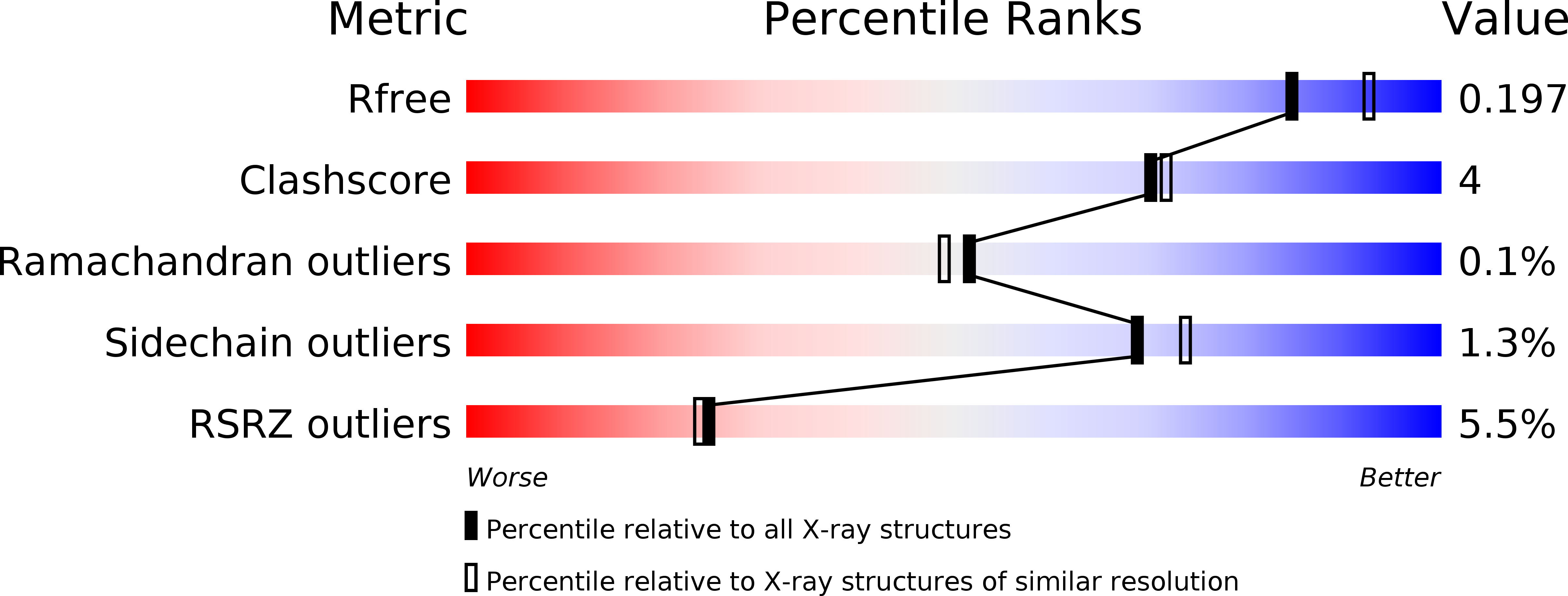
Resolution:
2.00 Å
R-Value Free:
0.19
R-Value Work:
0.16
R-Value Observed:
0.16
Space Group:
P 65
Macromolecular Entities
Polymer Type:polypeptide(L)
Molecule:Potassium voltage-gated channel subfamily H member 1
Gene (Uniprot):Kcnh1
Chain IDs:A, C, E, G
Chain Length:177
Number of Molecules:4
Biological Source:Mus musculus
Polymer Type:polypeptide(L)
Molecule:Potassium voltage-gated channel subfamily H member 1
Gene (Uniprot):Kcnh1
Chain IDs:B, D, F, H
Chain Length:134
Number of Molecules:4
Biological Source:Mus musculus
Primary Citation
The structural mechanism of KCNH-channel regulation by the eag domain.
Nature 501 444 448 (2013)
PMID: 23975098 DOI: 10.1038/nature12487

Abstact

The KCNH voltage-dependent potassium channels (ether-à-go-go, EAG; EAG-related gene, ERG; EAG-like channels, ELK) are important regulators of cellular excitability and have key roles in diseases such as cardiac long QT syndrome type 2 (LQT2), epilepsy, schizophrenia and cancer. The intracellular domains of KCNH channels are structurally distinct from other voltage-gated channels. The amino-terminal region contains an eag domain, which is composed of a Per-Arnt-Sim (PAS) domain and a PAS-cap domain, whereas the carboxy-terminal region contains a cyclic nucleotide-binding homology domain (CNBHD), which is connected to the pore through a C-linker domain. Many disease-causing mutations localize to these specialized intracellular domains, which underlie the unique gating and regulation of KCNH channels. It has been suggested that the eag domain may regulate the channel by interacting with either the S4-S5 linker or the CNBHD. Here we present a 2 Å resolution crystal structure of the eag domain-CNBHD complex of the mouse EAG1 (also known as KCNH1) channel. It displays extensive interactions between the eag domain and the CNBHD, indicating that the regulatory mechanism of the eag domain primarily involves the CNBHD. Notably, the structure reveals that a number of LQT2 mutations at homologous positions in human ERG, in addition to cancer-associated mutations in EAG channels, localize to the eag domain-CNBHD interface. Furthermore, mutations at the interface produced marked effects on channel gating, demonstrating the important physiological role of the eag domain-CNBHD interaction. Our structure of the eag domain-CNBHD complex of mouse EAG1 provides unique insights into the physiological and pathophysiological mechanisms of KCNH channels.

Legend

Protein

Chemical

Disease

Primary Citation of related structures