
Deposition Date
2013-07-04
Release Date
2013-08-28
Last Version Date
2024-11-13
Entry Detail
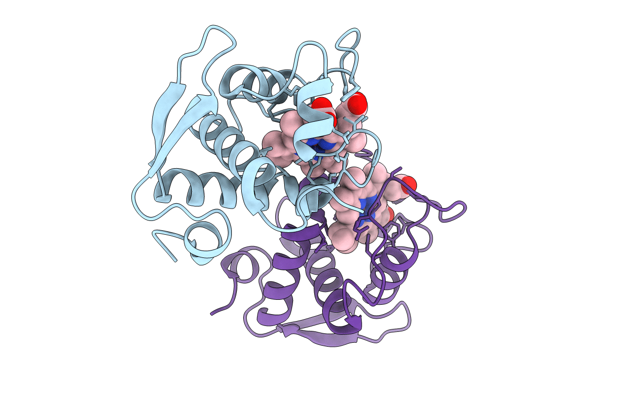
PDB ID:
4LJI
Keywords:
Title:
Crystal structure at 1.5 angstrom resolution of the PsbV2 cytochrome from the cyanobacterium thermosynechococcus elongatus
Biological Source:
Source Organism(s):
Thermosynechococcus elongatus (Taxon ID: 197221)
Method Details:
Experimental Method:
Resolution:
1.51 Å
R-Value Free:
0.25
R-Value Work:
0.21
R-Value Observed:
0.21
Space Group:
P 1
