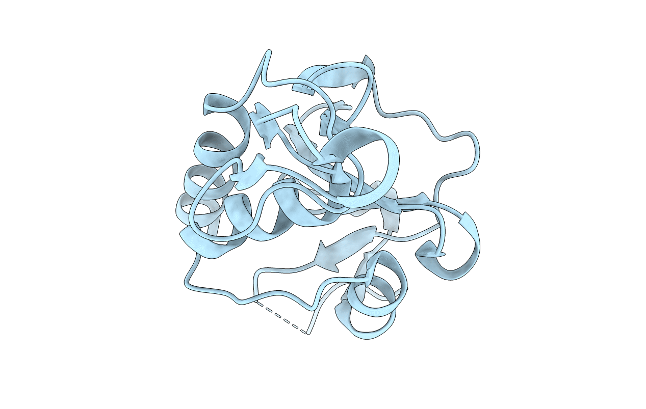
4LJ1 image
Entry Detail
PDB ID:
4LJ1
Keywords:
Title:
RipD (Rv1566c) from Mycobacterium tuberculosis: a non-catalytic NlpC/p60 domain protein with two penta-peptide repeat units (PVQQA-PVQPA)
Biological Source:
Host Organism:
PDB Version:
Deposition Date:
2013-07-04
Release Date:
2013-11-06
Method Details:
Experimental Method:
Resolution:
1.17 Å
R-Value Free:
0.16
R-Value Work:
0.14
R-Value Observed:
0.14
Space Group:
C 1 2 1
Macromolecular Entities
Polymer Type:polypeptide(L)
Description:Invasion-associated protein
Chain IDs:A
Chain Length:147
Number of Molecules:1
Biological Source:Mycobacterium tuberculosis
Primary Citation
RipD (Rv1566c) from Mycobacterium tuberculosis: adaptation of an NlpC/p60 domain to a non-catalytic peptidoglycan-binding function.
Biochem.J. 457 33 41 (2014)
PMID: 24107184 DOI: 10.1042/BJ20131227

Abstact

Enzymes carrying NlpC/p60 domains, for instance RipA and RipB from Mycobacterium tuberculosis, are bacterial peptidoglycan hydrolases that cleave the peptide stems and contribute to cell wall remodelling during cell division. A member of this protein family, RipD (Rv1566c) from M. tuberculosis described in the present study, displays sequence alterations in the NlpC/p60 catalytic triad and carries a pentapeptide repeat at its C-terminus. Bioinformatics analysis revealed RipD-like proteins in eleven mycobacterial genomes, whereas similar pentapeptide repeats occur in cell-wall-localized bacterial proteins and in a mycobacteriophage. In contrast with previously known members of the NlpC/p60 family, RipD does not show peptidoglycan hydrolase activity, which is consistent with the sequence alterations at the catalytic site. A strong interaction of the catalytically inactive core domain with peptidoglycan is however retained, presenting the first example of the NlpC/p60 domains that evolved to a non-catalytic peptidoglycan-binding function. Full-length RipD carrying the C-terminal repeat shows, however, a decrease in binding affinity to peptidoglycan, suggesting that the C-terminal tail modulates the interaction with bacterial cell wall components. The pentapeptide repeat at the C-terminus does not adopt a defined secondary structure in solution which is in accordance with results from the 1.17 Å (1 Å=0.1 nm) crystal structure of the protein carrying two repeat units.

Legend

Protein

Chemical

Disease

Primary Citation of related structures