4LGL image
Entry Detail
PDB ID:
4LGL
Keywords:
Title:
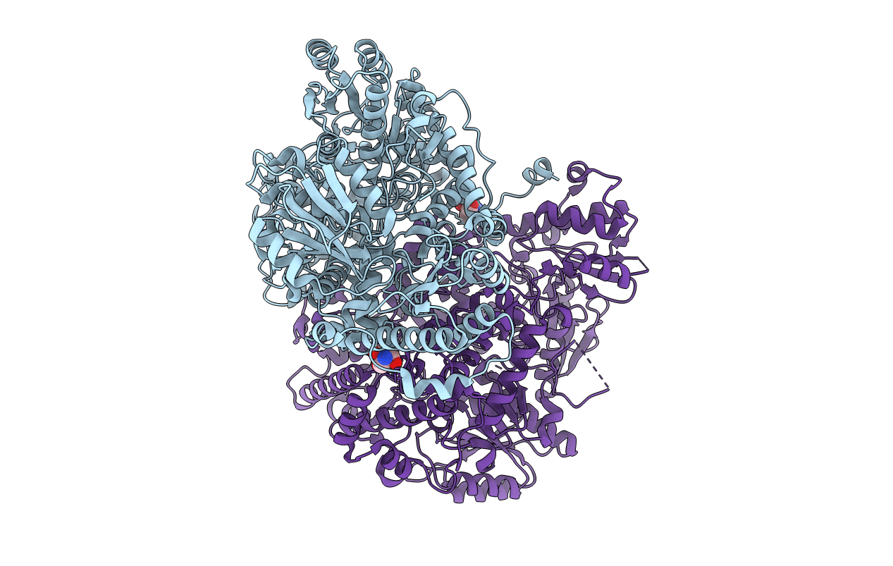
Crystal Structure of Glycine Decarboxylase P-protein from Synechocystis sp. PCC 6803, apo form
Biological Source:
Source Organism:
Host Organism:
PDB Version:
Deposition Date:
2013-06-28
Release Date:
2013-10-16
Method Details:
Experimental Method:
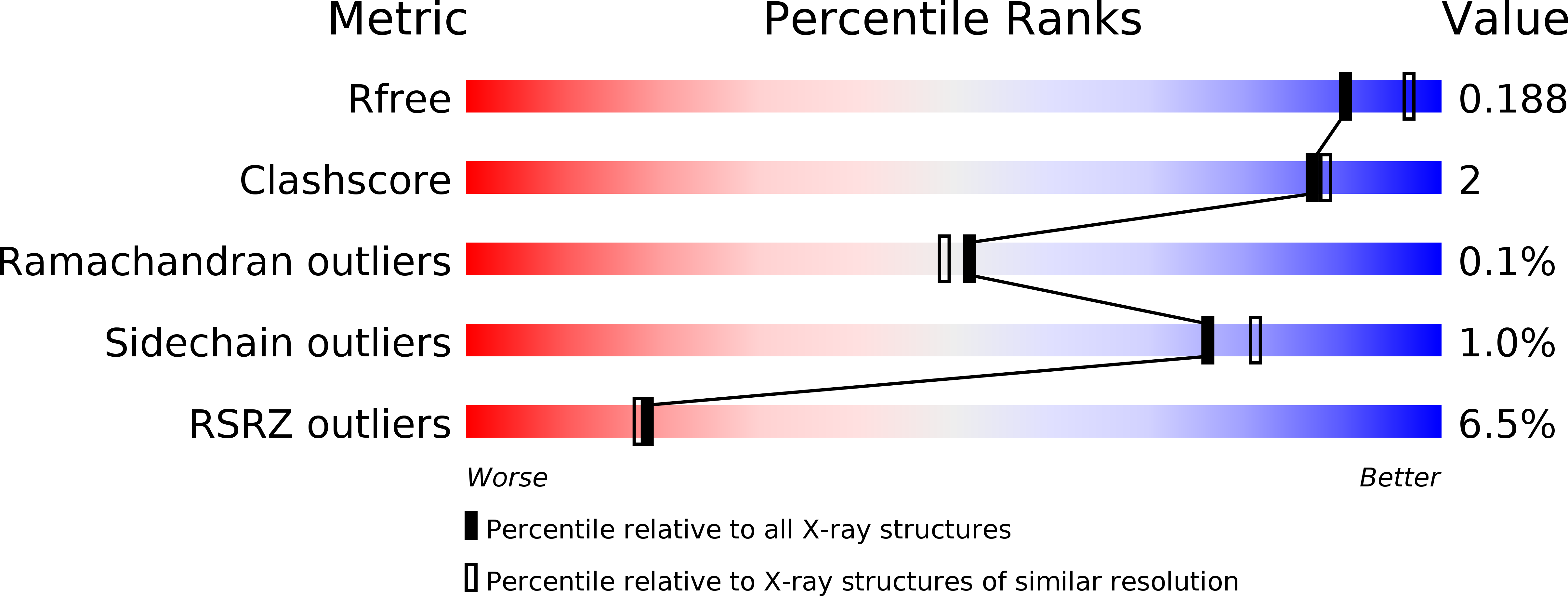
Resolution:
2.00 Å
R-Value Free:
0.19
R-Value Work:
0.16
R-Value Observed:
0.16
Space Group:
P 21 21 21
Macromolecular Entities
Polymer Type:polypeptide(L)
Description:Glycine dehydrogenase [decarboxylating]
Chain IDs:A, B
Chain Length:983
Number of Molecules:2
Biological Source:Synechocystis sp.
Modified Residue
Compound ID Chain ID Parent Comp ID Details 2D Image
CME A CYS S,S-(2-HYDROXYETHYL)THIOCYSTEINE
Primary Citation
Structure of the Homodimeric Glycine Decarboxylase P-protein from Synechocystis sp. PCC 6803 Suggests a Mechanism for Redox Regulation.
J.Biol.Chem. 288 35333 35345 (2013)
PMID: 24121504 DOI: 10.1074/jbc.M113.509976

Abstact

Glycine decarboxylase, or P-protein, is a pyridoxal 5'-phosphate (PLP)-dependent enzyme in one-carbon metabolism of all organisms, in the glycine and serine catabolism of vertebrates, and in the photorespiratory pathway of oxygenic phototrophs. P-protein from the cyanobacterium Synechocystis sp. PCC 6803 is an α2 homodimer with high homology to eukaryotic P-proteins. The crystal structure of the apoenzyme shows the C terminus locked in a closed conformation by a disulfide bond between Cys(972) in the C terminus and Cys(353) located in the active site. The presence of the disulfide bridge isolates the active site from solvent and hinders the binding of PLP and glycine in the active site. Variants produced by substitution of Cys(972) and Cys(353) by Ser using site-directed mutagenesis have distinctly lower specific activities, supporting the crucial role of these highly conserved redox-sensitive amino acid residues for P-protein activity. Reduction of the 353-972 disulfide releases the C terminus and allows access to the active site. PLP and the substrate glycine bind in the active site of this reduced enzyme and appear to cause further conformational changes involving a flexible surface loop. The observation of the disulfide bond that acts to stabilize the closed form suggests a molecular mechanism for the redox-dependent activation of glycine decarboxylase observed earlier.

Legend

Protein

Chemical

Disease

Primary Citation of related structures