4LFK image
Deposition Date 2013-06-27
Release Date 2013-11-27
Last Version Date 2023-11-08
Entry Detail
PDB ID:
4LFK
Keywords:
Title:
Crystal Structure of D-galactose-6-phosphate isomerase in a substrate-free form
Biological Source:
Source Organism:
Host Organism:
Method Details:
Experimental Method:
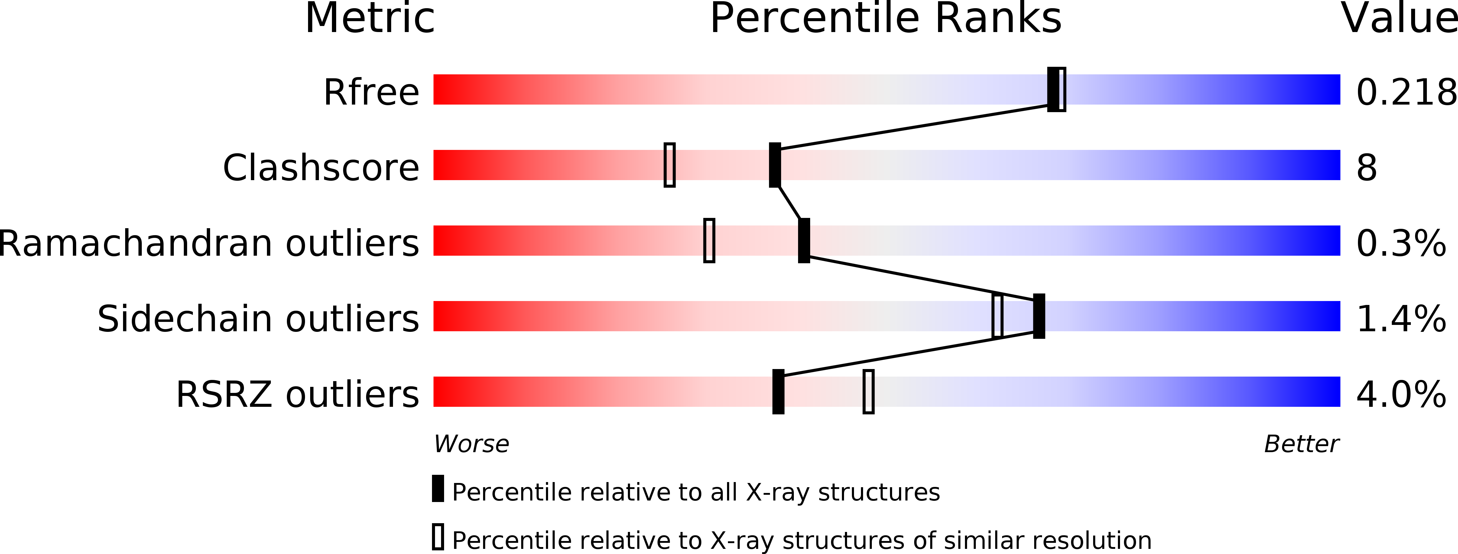
Resolution:
1.96 Å
R-Value Free:
0.21
R-Value Work:
0.20
Space Group:
P 21 21 2
Macromolecular Entities
Polymer Type:polypeptide(L)
Molecule:Galactose-6-phosphate isomerase subunit A
Chain IDs:A, C
Chain Length:162
Number of Molecules:2
Biological Source:Lactobacillus rhamnosus
Polymer Type:polypeptide(L)
Molecule:Galactose-6-phosphate isomerase subunit B
Chain IDs:B, D
Chain Length:172
Number of Molecules:2
Biological Source:Lactobacillus rhamnosus
Primary Citation
Crystal structure and substrate specificity of D-galactose-6-phosphate isomerase complexed with substrates.
Plos One 8 e72902 e72902 (2013)
PMID: 24015281 DOI: 10.1371/journal.pone.0072902

Abstact

D-Galactose-6-phosphate isomerase from Lactobacillus rhamnosus (LacAB; EC 5.3.1.26), which is encoded by the tagatose-6-phosphate pathway gene cluster (lacABCD), catalyzes the isomerization of D-galactose-6-phosphate to D-tagatose-6-phosphate during lactose catabolism and is used to produce rare sugars as low-calorie natural sweeteners. The crystal structures of LacAB and its complex with D-tagatose-6-phosphate revealed that LacAB is a homotetramer of LacA and LacB subunits, with a structure similar to that of ribose-5-phosphate isomerase (Rpi). Structurally, LacAB belongs to the RpiB/LacAB superfamily, having a Rossmann-like αβα sandwich fold as has been identified in pentose phosphate isomerase and hexose phosphate isomerase. In contrast to other family members, the LacB subunit also has a unique α7 helix in its C-terminus. One active site is distinctly located at the interface between LacA and LacB, whereas two active sites are present in RpiB. In the structure of the product complex, the phosphate group of D-tagatose-6-phosphate is bound to three arginine residues, including Arg-39, producing a different substrate orientation than that in RpiB, where the substrate binds at Asp-43. Due to the proximity of the Arg-134 residue and backbone Cα of the α6 helix in LacA to the last Asp-172 residue of LacB with a hydrogen bond, a six-carbon sugar-phosphate can bind in the larger pocket of LacAB, compared with RpiB. His-96 in the active site is important for ring opening and substrate orientation, and Cys-65 is essential for the isomerization activity of the enzyme. Two rare sugar substrates, D-psicose and D-ribulose, show optimal binding in the LacAB-substrate complex. These findings were supported by the results of LacA activity assays.

Legend

Protein

Chemical

Disease

Primary Citation of related structures