4LF2 image
Entry Detail
PDB ID:
4LF2
Title:
Hexameric Form II RuBisCO from Rhodopseudomonas palustris, activated and complexed with sulfate and magnesium
Biological Source:
Host Organism:
PDB Version:
Deposition Date:
2013-06-26
Release Date:
2014-06-25
Method Details:
Experimental Method:
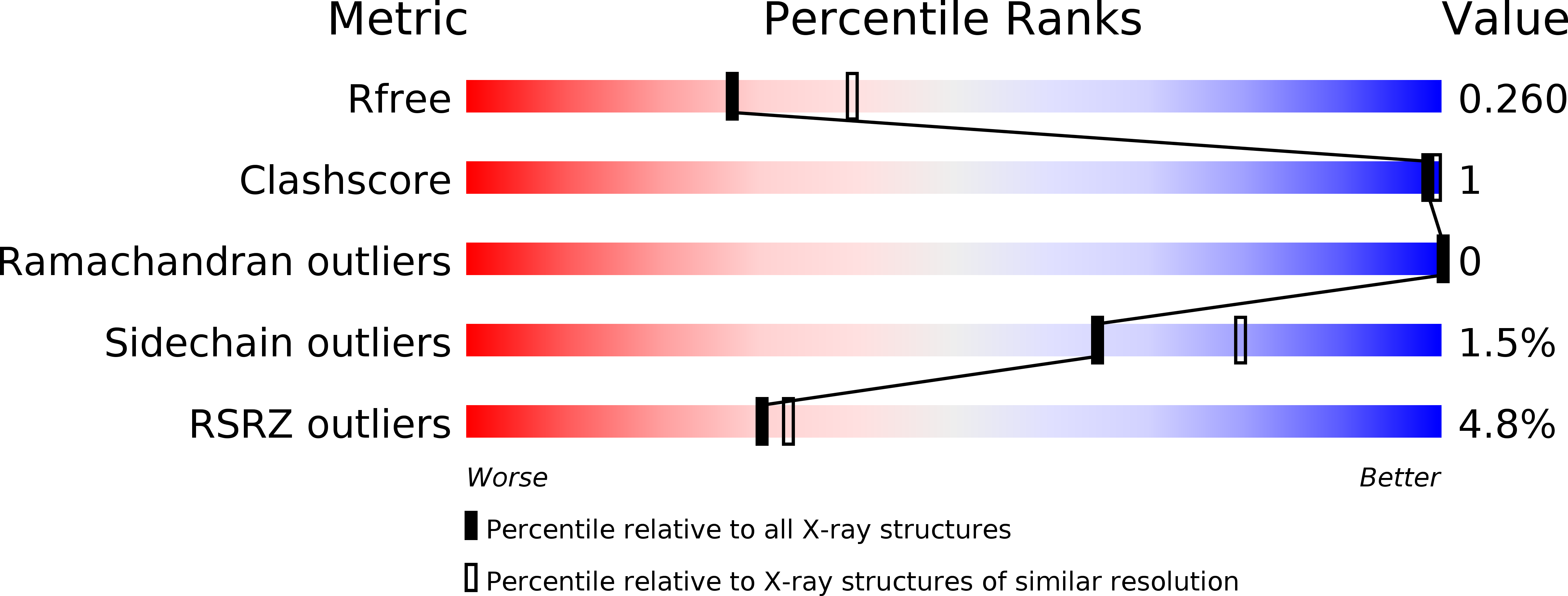
Resolution:
2.38 Å
R-Value Free:
0.25
R-Value Work:
0.20
R-Value Observed:
0.20
Space Group:
P 1
Macromolecular Entities
Polymer Type:polypeptide(L)
Description:Ribulose bisphosphate carboxylase
Chain IDs:A, B, C, D, E, F
Chain Length:481
Number of Molecules:6
Biological Source:Rhodopseudomonas palustris
Modified Residue
Compound ID Chain ID Parent Comp ID Details 2D Image
KCX A LYS LYSINE NZ-CARBOXYLIC ACID
Primary Citation
Structure-function studies with the unique hexameric form II ribulose-1,5-bisphosphate carboxylase/oxygenase (Rubisco) from Rhodopseudomonas palustris.
J.Biol.Chem. 289 21433 21450 (2014)
PMID: 24942737 DOI: 10.1074/jbc.M114.578625

Abstact

The first x-ray crystal structure has been solved for an activated transition-state analog-bound form II ribulose-1,5-bisphosphate carboxylase/oxygenase (Rubisco). This enzyme, from Rhodopseudomonas palustris, assembles as a unique hexamer with three pairs of catalytic large subunit homodimers around a central 3-fold symmetry axis. This oligomer arrangement is unique among all known Rubisco structures, including the form II homolog from Rhodospirillum rubrum. The presence of a transition-state analog in the active site locked the activated enzyme in a "closed" conformation and revealed the positions of critical active site residues during catalysis. Functional roles of two form II-specific residues (Ile(165) and Met(331)) near the active site were examined via site-directed mutagenesis. Substitutions at these residues affect function but not the ability of the enzyme to assemble. Random mutagenesis and suppressor selection in a Rubisco deletion strain of Rhodobacter capsulatus identified a residue in the amino terminus of one subunit (Ala(47)) that compensated for a negative change near the active site of a neighboring subunit. In addition, substitution of the native carboxyl-terminal sequence with the last few dissimilar residues from the related R. rubrum homolog increased the enzyme's kcat for carboxylation. However, replacement of a longer carboxyl-terminal sequence with termini from either a form III or a form I enzyme, which varied both in length and sequence, resulted in complete loss of function. From these studies, it is evident that a number of subtle interactions near the active site and the carboxyl terminus account for functional differences between the different forms of Rubiscos found in nature.

Legend

Protein

Chemical

Disease

Primary Citation of related structures