Deposition Date
2013-06-21
Release Date
2013-08-14
Last Version Date
2024-02-28
Entry Detail
PDB ID:
4LCD
Keywords:
Title:
Structure of an Rsp5xUbxSna3 complex: Mechanism of ubiquitin ligation and lysine prioritization by a HECT E3
Biological Source:
Source Organism(s):
Saccharomyces cerevisiae (Taxon ID: 559292)
Homo sapiens (Taxon ID: 9606)
Homo sapiens (Taxon ID: 9606)
Expression System(s):
Method Details:
Experimental Method:
Resolution:
3.10 Å
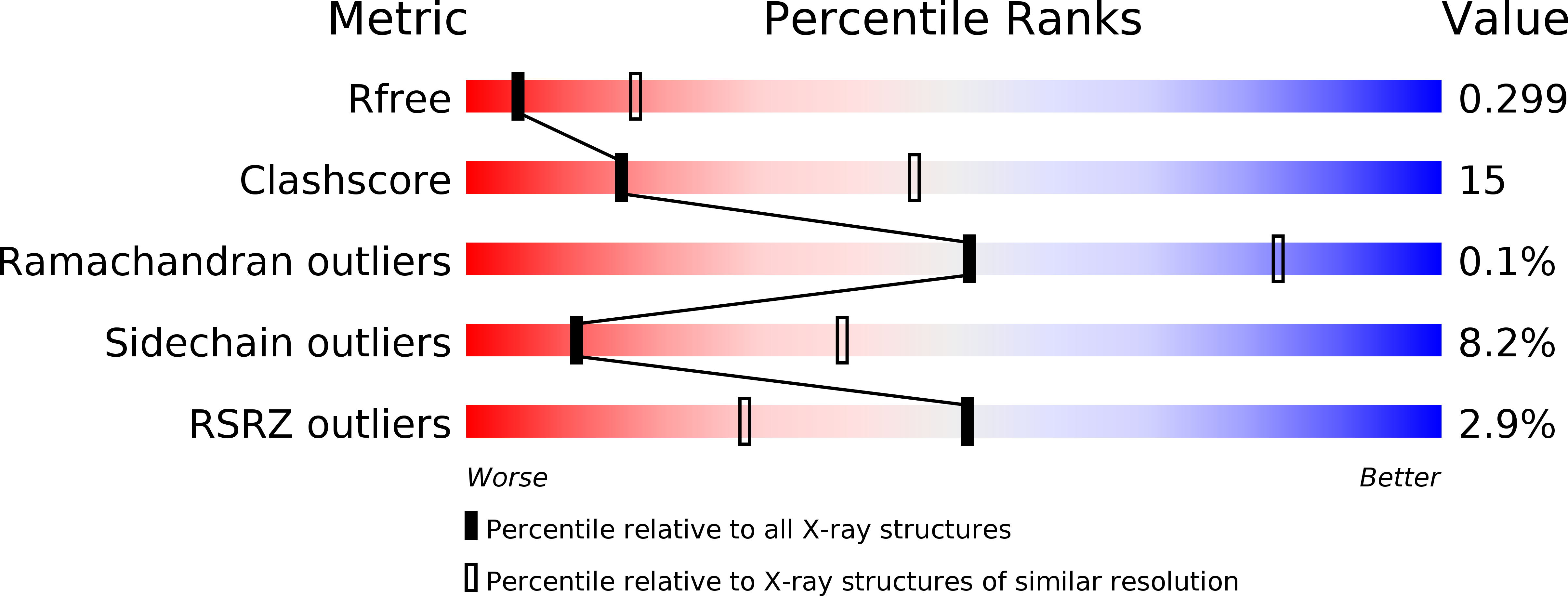
R-Value Free:
0.29
R-Value Work:
0.25
R-Value Observed:
0.25
Space Group:
P 1 21 1