4LB5 image
Deposition Date 2013-06-20
Release Date 2013-09-18
Last Version Date 2024-02-28
Entry Detail
PDB ID:
4LB5
Keywords:
Title:
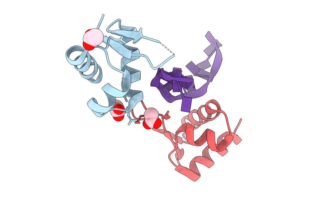
Crystal structure of PKZ Zalpha in complex with ds(CG)6 (hexagonal form)
Biological Source:
Source Organism:
Danio rerio (Taxon ID: 7955)
Host Organism:
Method Details:
Experimental Method:
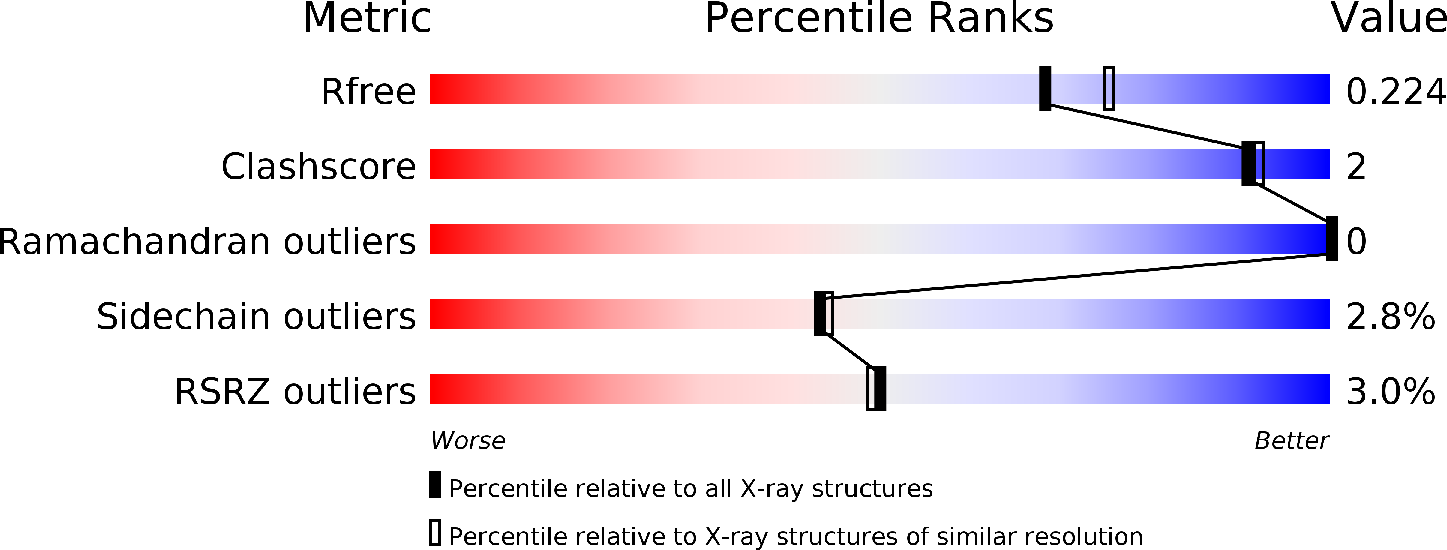
Resolution:
2.00 Å
R-Value Free:
0.22
R-Value Work:
0.18
R-Value Observed:
0.19
Space Group:
P 61 2 2
Macromolecular Entities
Structures with similar UniProt ID
Protein Blast
Polymer Type:polypeptide(L)
Molecule:Protein kinase containing Z-DNA binding domains
Gene (Uniprot):pkz
Chain IDs:A, B
Chain Length:72
Number of Molecules:2
Biological Source:Danio rerio
Polymer Type:polydeoxyribonucleotide
Molecule:5'-D(*TP*CP*GP*CP*GP*CP*GP*CP*GP*CP*GP*CP*G)-3'
Chain IDs:C
Chain Length:13
Number of Molecules:1
Biological Source:
Ligand Molecules
Primary Citation
Structural basis for Z-DNA binding and stabilization by the zebrafish Z-DNA dependent protein kinase PKZ.
Nucleic Acids Res. 41 9924 9933 (2013)
PMID: 23975196 DOI: 10.1093/nar/gkt743

Abstact

The RNA-dependent protein kinase PKR plays a central role in the antiviral defense of vertebrates by shutting down protein translation upon detection of viral dsRNA in the cytoplasm. In some teleost fish, PKZ, a homolog of PKR, performs the same function, but surprisingly, instead of dsRNA binding domains, it harbors two Z-DNA/Z-RNA-binding domains belonging to the Zalpha domain family. Zalpha domains have also been found in other proteins, which have key roles in the regulation of interferon responses such as ADAR1 and DNA-dependent activator of IFN-regulatory factors (DAI) and in viral proteins involved in immune response evasion such as the poxviral E3L and the Cyprinid Herpesvirus 3 ORF112. The underlying mechanism of nucleic acids binding and stabilization by Zalpha domains is still unclear. Here, we present two crystal structures of the zebrafish PKZ Zalpha domain (DrZalpha(PKZ)) in alternatively organized complexes with a (CG)6 DNA oligonucleotide at 2 and 1.8 Å resolution. These structures reveal novel aspects of the Zalpha interaction with DNA, and they give insights on the arrangement of multiple Zalpha domains on DNA helices longer than the minimal binding site.

Legend

Protein

Chemical

Disease

Primary Citation of related structures
Feedback Form
Name
Email
Institute
Feedback