4L8S image
Deposition Date 2013-06-17
Release Date 2013-10-16
Last Version Date 2025-03-26
Entry Detail
PDB ID:
4L8S
Keywords:
Title:
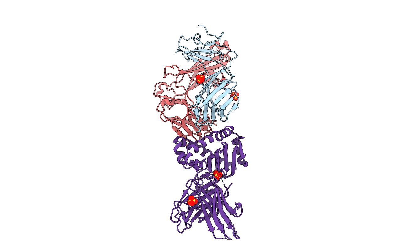
Crystal structure of a human Valpha7.2/Vbeta13.3 MAIT TCR in complex with bovine MR1
Biological Source:
Source Organism:
Homo sapiens (Taxon ID: 9606)
Bos taurus (Taxon ID: 9913)
Host Organism:
Method Details:
Experimental Method:
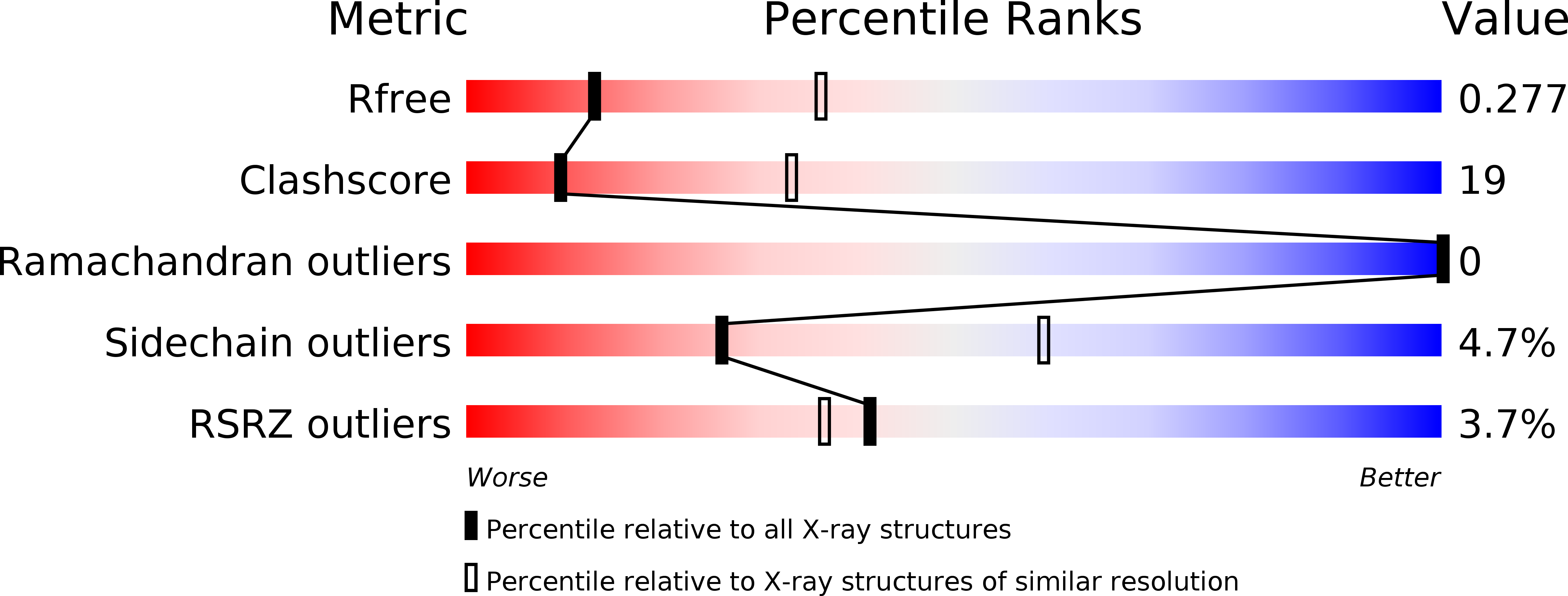
Resolution:
2.90 Å
R-Value Free:
0.27
R-Value Work:
0.22
R-Value Observed:
0.23
Space Group:
P 21 21 21
Macromolecular Entities
Polymer Type:polypeptide(L)
Molecule:Muccosal Associated Invariant T Cell Receptor alpha chain
Mutations:T157C
Chain IDs:A
Chain Length:208
Number of Molecules:1
Biological Source:Homo sapiens
Polymer Type:polypeptide(L)
Molecule:Muccosal Associated Invariant T Cell Receptor beta chain
Mutations:S172C
Chain IDs:B
Chain Length:252
Number of Molecules:1
Biological Source:Homo sapiens
Polymer Type:polypeptide(L)
Molecule:Beta-2-microglobulin, MHC class I-related protein
Gene (Uniprot):MR1, B2M
Chain IDs:C
Chain Length:392
Number of Molecules:1
Biological Source:Bos taurus
Modified Residue
Compound ID Chain ID Parent Comp ID Details 2D Image
KFP C LYS ?
Ligand Molecules
Primary Citation
MAIT Recognition of a Stimulatory Bacterial Antigen Bound to MR1.
J.Immunol. 191 5268 5277 (2013)
PMID: 24108697 DOI: 10.4049/jimmunol.1301958

Abstact

MR1-restricted mucosal-associated invariant T (MAIT) cells represent a subpopulation of αβ T cells with innate-like properties and limited TCR diversity. MAIT cells are of interest because of their reactivity against bacterial and yeast species, suggesting that they play a role in defense against pathogenic microbes. Despite the advances in understanding MAIT cell biology, the molecular and structural basis behind their ability to detect MR1-Ag complexes is unclear. In this study, we present our structural and biochemical characterization of MAIT TCR engagement of MR1 presenting an Escherichia coli-derived stimulatory ligand, rRL-6-CH2OH, previously found in Salmonella typhimurium. We show a clear enhancement of MAIT TCR binding to MR1 due to the presentation of this ligand. Our structure of a MAIT TCR/MR1/rRL-6-CH2OH complex shows an evolutionarily conserved binding orientation, with a clear role for both the CDR3α and CDR3β loops in recognizing the rRL-6-CH2OH stimulatory ligand. We also present two additional xenoreactive MAIT TCR/MR1 complexes that recapitulate the docking orientation documented previously, despite having variation in the CDR2β and CDR3β loop sequences. Our data support a model by which MAIT TCRs engage MR1 in a conserved fashion, with their binding affinities modulated by the nature of the MR1-presented Ag or diversity introduced by alternate Vβ usage or CDR3β sequences.

Legend

Protein

Chemical

Disease

Primary Citation of related structures