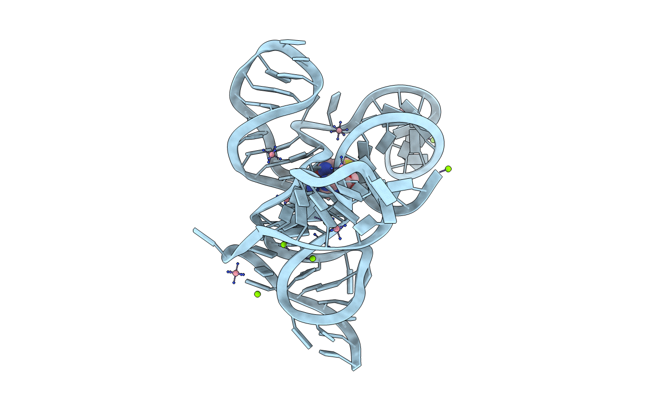
4L81 image
Entry Detail
PDB ID:
4L81
Keywords:
Title:
Structure of the SAM-I/IV riboswitch (env87(deltaU92, deltaG93))
Biological Source:
Source Organism:
PDB Version:
Deposition Date:
2013-06-15
Release Date:
2014-05-28
Method Details:
Experimental Method:
Resolution:
2.95 Å
R-Value Free:
0.23
R-Value Work:
0.21
R-Value Observed:
0.21
Space Group:
P 64 2 2
Macromolecular Entities
Polymer Type:polyribonucleotide
Description:SAM-I/IV variant riboswitch aptamer domain
Chain IDs:A
Chain Length:96
Number of Molecules:1
Biological Source:
Primary Citation
Structural basis for diversity in the SAM clan of riboswitches.
Proc.Natl.Acad.Sci.USA 111 6624 6629 (2014)
PMID: 24753586 DOI: 10.1073/pnas.1312918111

Abstact

In bacteria, sulfur metabolism is regulated in part by seven known families of riboswitches that bind S-adenosyl-l-methionine (SAM). Direct binding of SAM to these mRNA regulatory elements governs a downstream secondary structural switch that communicates with the transcriptional and/or translational expression machinery. The most widely distributed SAM-binding riboswitches belong to the SAM clan, comprising three families that share a common SAM-binding core but differ radically in their peripheral architecture. Although the structure of the SAM-I member of this clan has been extensively studied, how the alternative peripheral architecture of the other families supports the common SAM-binding core remains unknown. We have therefore solved the X-ray structure of a member of the SAM-I/IV family containing the alternative "PK-2" subdomain shared with the SAM-IV family. This structure reveals that this subdomain forms extensive interactions with the helix housing the SAM-binding pocket, including a highly unusual mode of helix packing in which two helices pack in a perpendicular fashion. Biochemical and genetic analysis of this RNA reveals that SAM binding induces many of these interactions, including stabilization of a pseudoknot that is part of the regulatory switch. Despite strong structural similarity between the cores of SAM-I and SAM-I/IV members, a phylogenetic analysis of sequences does not indicate that they derive from a common ancestor.

Legend

Protein

Chemical

Disease

Primary Citation of related structures