
Deposition Date
2013-06-14
Release Date
2014-06-18
Last Version Date
2023-11-08
Entry Detail
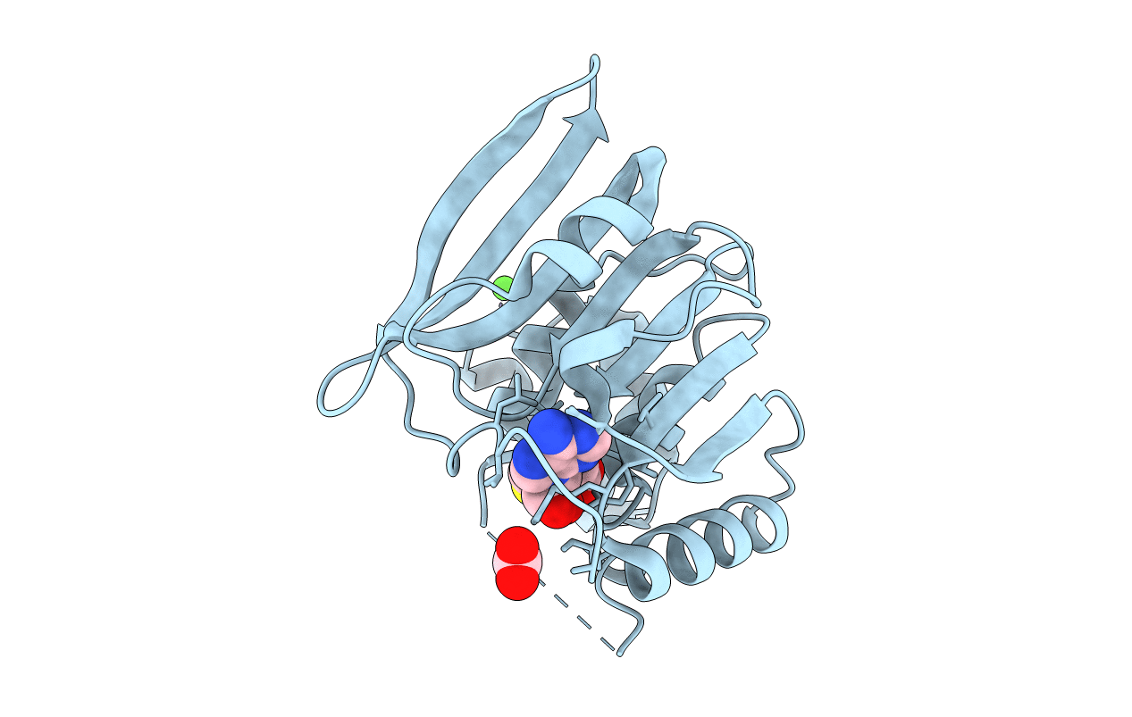
PDB ID:
4L7V
Keywords:
Title:
Crystal structure of Protein L-isoaspartyl-O-methyltransferase of Vibrio cholerae
Biological Source:
Source Organism(s):
Vibrio cholerae (Taxon ID: 345073)
Expression System(s):
Method Details:
Experimental Method:
Resolution:
2.05 Å
R-Value Free:
0.27
R-Value Work:
0.23
R-Value Observed:
0.23
Space Group:
P 41 21 2
