4L7B image
Entry Detail
PDB ID:
4L7B
Title:
Structure of keap1 kelch domain with (1S,2R)-2-{[(1S)-1-[(1,3-dioxo-1,3-dihydro-2H-isoindol-2-yl)methyl]-3,4-dihydroisoquinolin-2(1H)-yl]carbonyl}cyclohexanecarboxylic acid
Biological Source:
Source Organism:
Host Organism:
PDB Version:
Deposition Date:
2013-06-13
Release Date:
2014-02-19
Method Details:
Experimental Method:
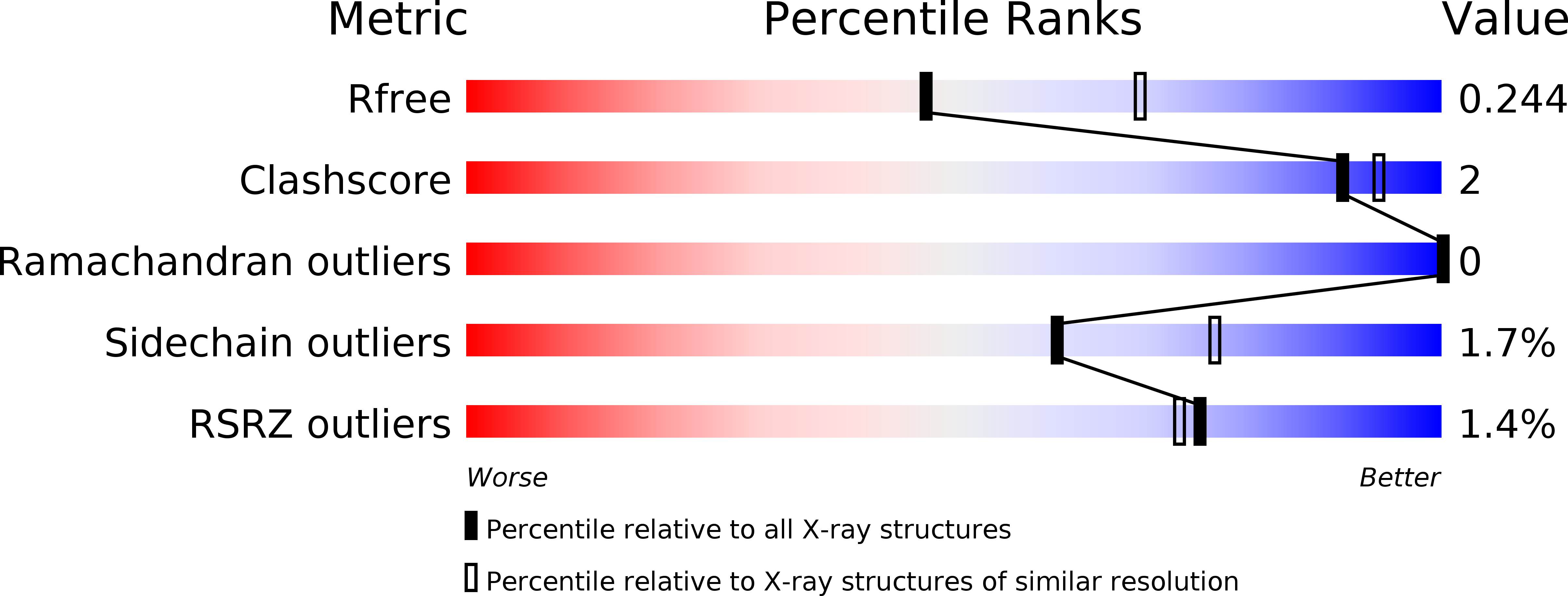
Resolution:
2.41 Å
R-Value Free:
0.24
R-Value Work:
0.22
R-Value Observed:
0.22
Space Group:
P 21 21 21
Macromolecular Entities
Polymer Type:polypeptide(L)
Description:Kelch-like ECH-associated protein 1
Mutations:R354D
Chain IDs:A, B
Chain Length:300
Number of Molecules:2
Biological Source:Homo sapiens
Primary Citation
Binding Mode and Structure-Activity Relationships around Direct Inhibitors of the Nrf2-Keap1 Complex.
Chemmedchem 9 699 705 (2014)
PMID: 24504667 DOI: 10.1002/cmdc.201300525

Abstact

An X-ray crystal structure of Kelch-like ECH-associated protein (Keap1) co-crystallised with (1S,2R)-2-[(1S)-1-[(1,3-dioxo-2,3-dihydro-1H-isoindol-2-yl)methyl]-1,2,3,4-tetrahydroisoquinolin-2-carbonyl]cyclohexane-1-carboxylic acid (compound (S,R,S)-1 a) was obtained. This X-ray crystal structure provides breakthrough experimental evidence for the true binding mode of the hit compound (S,R,S)-1 a, as the ligand orientation was found to differ from that of the initial docking model, which was available at the start of the project. Crystallographic elucidation of this binding mode helped to focus and drive the drug design process more effectively and efficiently.

Legend

Protein

Chemical

Disease

Primary Citation of related structures